Single Ventricle Heart Defect and Treatment Explained - DILV
What is a single ventricle congenital heart defect and how is it treated? Parents of a newborn with a heart defect called Double-Inlet Left Ventricle (DILV) use simple illustrations to explain what this particular single ventricle defect is, how it affects the child's health, and what the surgical treatment will look like.
Parents John and Megan Filson talk about the emotional impact of their baby's heart condition and the complex surgeries required to address it.
The CDC estimates nearly one percent of babies born in the United States are affected by some form of congenital heart defect. Some common defects include: holes between the heart's chambers (ventricular and atrial septal defects, Tetraology of Fallot), misplaced, blocked, or malfunctioning arteries or valves (transposition, stenosis, Truncus Arteriosus, PDA, Ebstein's Anomaly), and single ventricle (Hypoplastic Left Heart, Double-Inlet Left Ventricle (DILV), Double-Outlet Right Ventricle (DORV))
Babies born with a single ventricle heart defect face the life-threatening problem of not getting enough blood flow to their lungs, or getting too much blood flow that causes fatal damage to the lungs. They also face the problem of not getting enough oxygen to their body.
A Partial Norwood, or Damus-Kaye-Stansel procedure, attaches the pulmonary artery to the aorta and uses a synthetic shunt to pass blood from the aorta to the lungs.
The two-stage Fontan operation connects the baby's vena cava to the pulmonary arteries in order to bypass the heart and prevent oxygenated blood from getting mixed with de-oxygenated blood.
The first stage is called the Hemi Fontan or the Glenn Procedure, performed when the baby is about 6 months old. It connects the superior vena cava directly to the pulmonary arteries.
The second stage is the Fontan Procedure, performed when the child is about three years old. It connects the inferior vena cava to the pulmonary arteries.
After these surgeries, single ventricle children can live a normal life. There will be strain placed on the liver, kidneys, lungs and heart that may require major interventions later in life.
Helpful resources:
Children's Hospital Los Angeles:
www.chla.congenital.org
American Heart Association:
www.heart.org - conditions - congenital defects
Mended Little Hearts:
https://www.facebook.com/MendedLittleHeartsNationalOrganization
Видео Single Ventricle Heart Defect and Treatment Explained - DILV канала John Filson
Показать
Комментарии отсутствуют
Информация о видео
Другие видео канала
 Single Ventricle: Life After Surgery - The Children's Hospital of Philadelphia (6 of 6)
 Babies with misshapen hearts: single-ventricle defects
 Hypoplastic left heart syndrome and norwood glenn fontan | NCLEX-RN | Khan Academy
 What It's Like to Be an Adult with a Congenital Heart Defect
 Baby circulation right after birth | Circulatory system physiology | NCLEX-RN | Khan Academy
 Tetralogy of Fallot (TOF): Animation Explains Heart Defect and Repair
 Transposition of the Great Arteries (Vessels) | Congenital Heart Defects Nursing NCLEX Pediatrics
 Single Ventricle Home Monitoring Program
 Single Ventricle 1: Anatomy/Physiology and Pre-Fontan Palliation (Thomas Young, MD)
 Left sided vs. Right sided heart failure
 HLHS & Norwood-Glenn-Fontan
![[7 minutes] Single Ventricle Physiology MADE CLEAR by Pediatric Cardiology PLAYBOOK. Explained](https://i.ytimg.com/vi/GdClYvQQ9VI/default.jpg) [7 minutes] Single Ventricle Physiology MADE CLEAR by Pediatric Cardiology PLAYBOOK. Explained
[7 minutes] Single Ventricle Physiology MADE CLEAR by Pediatric Cardiology PLAYBOOK. Explained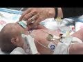 Single Ventricle Malformations - The Children's Hospital of Philadelphia (1 of 6)
 Assessing Right Ventricular Function - ECHO Course l The EKG Guy - www.ekg.md
 Introduction & Chapter- 1 of 24 Systematic approach to Segmental analysis of CHD
 Single Ventricle 1: Anatomy/Physiology and Pre-Fontan Palliation (THOMAS W. YOUNG, MD)
 Double Outlet Right Ventricle
 Development of Heart Tube | Heart Embryology
 "Ventricular Septal Defects" by Dr. David Bailly for OPENPediatrics
 Single Ventricle Surgery: Norwood Procedure - The Children's Hospital of Philadelphia (3 of 6)