Acute Pulmonary Embolism
Several signs that can be detected by echocardiography are suggestive of acute Pulmonary Embolism (PE), including right ventricle (RV) hypokinesia, McConnell’s sign, pulmonary artery hypertension, RV tissue Doppler, TAPSE, RV thrombus and tricuspid regurgitation. * However, when a patient presents with a submassive or larger pulmonary embolism, by definition RV dysfunction will be present on the echo. Our patient presented with a shock index greater than 1 (HR/SBP), suggesting a potentially large clot.
* One echo finding that has been found to be 94% specific for PE is the so called 60/60 sign. This refers to an RV systolic pressure (RVSP) less than 60 mmHg and a pulmonary acceleration time (PAT) less than 60 msec.
* A proximal occlusion (PE) causes the velocity to peak quickly with (PAT) little vascular compliance: imagine blowing through an occluded straw.
* RV systolic Tissue Doppler less than 10 cm/s is abnormal and should be included with evaluating PE. This patient was 4 cm/s.
* TAPSE measurement during m-mode evaluation was significantly reduced measuring 7 cm/s. Normal value is 17 or above.
* If there is right ventricular free wall hypokinesis in the presence of normal right ventricular apical contractility, this is known as McConnell Sign. Please keep in mind ***Mcconnell Sign has a 94% specificity for PE, but subsequent studies have shown that it can be seen in acute RV infarct and up to 17% of patients with chronic pulmonary hypertension.
Go follow my IG: @ultrasound_connection
#PulmonaryEmbolism #Cardiology #EmergencyMedicine #InternalMedicine #Hospitalist #FamilyMedicine #Anesthesiology #NursePractitioner #PA #cardiology #echocardiography #sonographersdoitinthedark #ultrasoundtech #ultrasoundtechniques #ultrasoundtechnician
Видео Acute Pulmonary Embolism канала Ultra Girl
Показать
Комментарии отсутствуют
Информация о видео
Другие видео канала
 Abnormal Strain Echocardiogram
 How to clean 🧼 TEE Probe after procedure 🤩
 Abnormal RV Strain| Acute RV Syndrome 💔 #echocardiography #RVstrain #shorts #medicine #echo
 Transcranial Doppler (Part 3) Transorbital View #science #echocardiography #medicine #shorts
 Ultrasound findings for dehydration #echocardiogram
 The Best Ultrasound Escape 🤍😍| Heart Tingz #echocardiography #medicine #science #techlife
 Left Atrial Myxoma 🫀😰
 Coronary artery disease 🫀
 3D TEE of the mitral valve LA/LV 🧡💛
 Vascular health! 💪🏥 Here’s to reduced stroke risk and a healthier future! 🙌
 Surgery complications TAVR #shorts #echocardiography #exploremore
 Echo Trivia Time #Cardioversion 💓
 How to do Ankle Brachial Index for arterial disease |ABI Ratio
 Cardiovascular drugs their use and side effects 💊 #echocardiography #sonography
 Tips for obtaining standard Echo views and heart anatomy 🤪🫶🏽
 Myocardial infarction with latent aneurysm ventricular septal defect 🫣💔
 Tricuspid Valve Vegetation 😱 #echocardiogram #vegetation #ultrasound #sonography
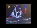 How to do strain echocardiogram ❤️✨
 Carotid Artery Anatomy (Part 1 of Transcranial Doppler) #echocardiography #science #sonography
 Echo (Positive Bubble Study)