Nervous System Tumors
This is a summary of the major tumors of the nervous system.
I created this presentation with Google Slides.
Image were created or taken from Wikimedia Commons
I created this video with the YouTube Video Editor.
ADDITIONAL TAGS:
Medulloblastoma
Pilocytic astrocytoma
Diffuse astrocytoma
Anaplastic astrocytoma
Glioblastoma
Oligodendrogliomas
Ependymomas
Meningiomas
Neurofibromas
Schwannomas
By James Heilman, MD - Own work, CC BY-SA 3.0, https://commons.wikimedia.org/w/index.php?curid=16272952
Embryonal tumor
Meningeal tumor
Peripheral tumor
Astrocytoma
Gliomas
Medulloblastoma
Grade IV malignant tumor
Arises from external granular layer in cerebellum
Histo: small round blue cells; Homer-Wright rosettes; mitotically active
Located in the cerebellum
Can drop metastases to seed the CSF
Well-circumscribed (non diffuse, solid)
By Jensflorian - Own work, CC BY-SA 3.0, https://commons.wikimedia.org/wiki/File:Neuroblastoma_Homer_Wright_rosettes_HE.jpg
Pilocytic astrocytoma
Grade I
Imaging: Solid nodule with cystic component
Common in cerebellum, hypothalamus, and optic nerve
Well-circumscribed (non diffuse)
Histo: Rosenthal fibers, eosinophilic granular bodies
BRAF mutation
Tumor in hypthalmic region - By The Armed Forces Institute of Pathology - http://peir2.path.uab.edu/scripts/acdis.dll?cmd=see&fp=/dbih/AFIP/00405615.tif&fmt=jpg&q=100&h=512, Public Domain, https://commons.wikimedia.org/w/index.php?curid=6182037
ROSENTHAL FIBERS - By Marvin 101 - Own work, CC BY-SA 3.0, https://commons.wikimedia.org/w/index.php?curid=5766818
Diffuse and anaplastic astrocytoma
Diffuse astrocytoma
Grade II malignant tumor
Diffuse
Most common in cerebral hemispheres
Histo: hypercellular
Molecular: no 1p/19p codeletion
Anaplastic astrocytoma
Grade III malignant tumor
Diffuse
Common location is cerebral hemispheres
Histo: hypercellular, increased mitotic activity
Molecular: no 1p/19p codeletion
Glioblastoma
Grade IV malignant tumor
Diffuse, infiltrative mass
Common location is cerebral hemispheres
Ring enhancement on imaging; internal necrosis
Histo: hypercellular, increased mitotic activity, microvascular proliferation, necrotizing, pseudopalisading
Molecular: EGFR, PTEN
Ring enhancing- By Christaras A - Created myself from anonymized patient MR, CC BY 2.5, https://commons.wikimedia.org/w/index.php?curid=1247038
Hypercellularity - By No machine-readable author provided. KGH assumed (based on copyright claims). - No machine-readable source provided. Own work assumed (based on copyright claims)., CC BY-SA 3.0, https://commons.wikimedia.org/w/index.php?curid=516831
Oligodendrogliomas
Grade II-III malignant tumor
Common location is cerebral hemispheres
Histo: hypercellular, perinuclear halo (fried-egg appearance), negative for astrocyte marker
Molecular: 1p/19q co-deletions
Perinuclear halo - By Nephron - Own work, CC BY-SA 3.0, https://commons.wikimedia.org/w/index.php?curid=7437415
Ependymomas
Grade II-III malignant tumor
Common in cerebellum and spinal cord
Well-circumscribed (non diffuse)
Can drop metastases to seed the CSF
Histo: perivascular rosettes, ependymal rosettes
Rosettes - By Nephron - Own work, CC BY-SA 3.0, https://commons.wikimedia.org/w/index.php?curid=10714428
meningiomas
Grade I benign tumor
Tumor of adults, not children
More common in females
Might respond to hormone growth (progesterone/estrogen receptors)
Dural tails; extra-axial location
Can invade bone and less-commonly brain
Histo: cellular whorls, psammoma bodies (calcium deposits)
Whorls - By Nephron - Own work, CC BY-SA 3.0, https://commons.wikimedia.org/w/index.php?curid=10728713
Dural tails
By Glitzy queen00 - http://en.wikipedia.org/wiki/Image:Contrast_enhanced_meningioma.jpg, Public Domain, https://commons.wikimedia.org/w/index.php?curid=2359385
Neurofibromas
Associated with neurofibromatosis type I
Cafe-au-lait spots, lisch nodules in eye
Histo: “shredded carrot appearance†of collagen strands
Lisch nodules - By Dimitrios Malamos - Own work, CC BY 4.0, https://commons.wikimedia.org/w/index.php?curid=45193921
Histo - By No machine-readable author provided. KGH assumed (based on copyright claims). - No machine-readable source provided. Own work assumed (based on copyright claims)., CC BY-SA 3.0, https://commons.wikimedia.org/w/index.php?curid=443285
Schwannomas
Can be associated with NF2
Histo:
verocay bodies (palisading cells surrounding acellular zone)
biphasic neoplasm with Antoni A (hypercellular) and Antoni B (hypocellular) regions
Common: Vestibular schwannoma (aka acoustic neuroma) from CN VIII
Hearing problems and disequilibrium
Medulloblastoma
Pilocytic astrocytoma
Diffuse astrocytoma
Anaplastic astrocytoma
Glioblastoma
Oligodendrogliomas
Ependymomas
Meningiomas
Neurofibromas
Schwannomas
Biphasic histo - By Nephron - Own work, CC BY-SA 3.0, https://commons.wikimedia.org/w/index.php?curid=17748282
Видео Nervous System Tumors канала MedLecturesMadeEasy
Показать
Комментарии отсутствуют
Информация о видео
Другие видео канала
 Meningiomas - Dr. Isaac Yang | UCLA Neurosurgery
 Brain Tumors
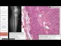 Bone and cartilage tumors
 Tumors of the Central Nervous System - CRASH! Medical Review Series
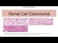 Renal cell carcinoma: Types,Genetics,Morphology
 Growths, neoplasms, and cancers of the skin
 Histopathology Brain--Astrocytoma
 Brain herniation - causes, symptoms, diagnosis, treatment, pathology
 Imaging of Brain Tumors
 Cognitive Changes in Patients with Brain Tumors | Memorial Sloan Kettering
 Malignant Peripheral Nerve Sheath Tumor (MPNST)...Explained by a Sarcoma Pathologist
 Pediatric Brain Tumors and Solid Masses
 Thyroid Cancers
 Adult brain tumors - causes, symptoms, diagnosis, treatment, pathology
 USMLE Neurology 24 Neuro Pathology: Brain Neoplasms and Cancer
 The Nervous System In 9 Minutes
 CNS Trauma
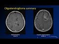 Imaging brain tumors - 3 - Oligodendrogliomas
 Multiple sclerosis - causes, symptoms, diagnosis, treatment, pathology
 Imaging brain tumors - 2 - Astrocytomas