Anatomy of the Heart - External and Internal Structures
This video is about the structural anatomy of the heart
Circulation of the heartCirculus Sanguid Minor (Pulmonary circulation):
- Deoxygenated blood comes from the body into the right atrium though superior and inferior vena cava.
- Blood goes through Tricuspid valve into right ventricle
- Right ventricle pumps blood through pulmonary valve into pulmonary artery
- Pulmonary artery pumps blood into the lungs
Circulus Sanguis major (Systemic Circulation):
- Blood comes from the lungs into the left Atria
- Left atria sends blood through bicuspid valve into the left ventricle.
- Left ventricle sends blood through the Aortic Valve into the Aorta
- Aorta sends blood to the body again
External Structures of the heart
- Apex Cordis directed anteriorly to the left
- Basis Cordis directed backwards to the right.
Three surfaces fo the heart:
- Facies pulmonalis
- Facies Sternocostalis
- Facies Diaphragmatica
Strictures on the surface of the heart:
- Margo Dexter between Facies Sternocostalis and Facies Diaphragmatica
- Sulcus Coronarius separates Atria from Ventricles
- Sulcus Interventricularis anterior and posterior.
Internal Septum:
- Septum Cordis separates right side from left side.
- Septum interventriculare (Muscular part and Membranous part)
- Septum Interatriale
Internal structures of the heart
Walls of the Right Atrium (Strictures):
- Anterior Wall: Auricula Dextra (Pectinate Muscle)
- Lateral Wall: Musculus Pectinati
- Superior Wall: Ostium Vena Cava Superior
- Between Superior and Inferior Vena Cava is the Sinus Venarum Cavarum (Sinus Venosum) separated from pectinate muscle by a crest on outside called Crista Terminalis
- Posterior Wall: Ostrium Vena Cava Inferior, Vulva vena cava inferioris (Inferior Vena Caval valve)
- Medial Wall: Septum Interatriale, Fossa Ovale (Foramen Ovale) Limbus Fossa Ovale, Ostium Sinus Coronarii, Vulva SInus Coronarii, Foramina Venarum Minimarum.
- Inferior Wall: Ostium Atrioventriculare Dextrum, guarded by the tricuspid valve.
Strictures of the Right Ventricle:
- Rough inner layer; Trabeculae Carneae
- Musculi Papillares
Base of the right ventricle:
Tricuspid Valve: Septal Cusp, Anterior cusp and posterior cusp formed by endocardium
- Surrounded by anulus fibrosus
- Chordae Tendinae attached to musculi pappilares.
Pulmonary Valve:
- Also has Anulus Fibrosus
- Has Noduli Vulva Semilunaris (Nodules of the Semilunar valve)
- Sinus Trunci Pulmonares (Pulmonary Sinus)
- Three Semilunar Cusps
Walls of the Left Atrium:
- Anterior Wall: Auricula Sinister (Has Musculi Pectinati)
- Posterior Wall: Pulmonary Veins (has 4 openings called ostia ventarum pulmonalium, openings of the pulmonary veins)
- Medial Wall: Septa interatriale
- Inferior Wall: Ostium Atrioventriculare sinister
Structures of the left ventricle:
Base of the ventricle:
- Ostium Atrioventriculare Sinister guarded by Bicuspid Valve (Mitral Valve)
- Bicuspid Valve has two big cusps or leaflets called cuspis anterior and cuspis posterior. But it also has Commissural Cusps.
- Ostium Aorta (in Vestibulum Aortae)
- Vulva Aortae (three semilunar valve)
- Has Aortic Sinuses and Aortic Nodules
- Trabeculae Carneae
- Musculi Papillares
Hi:)
Meditay
Видео Anatomy of the Heart - External and Internal Structures канала Meditay
Показать
Комментарии отсутствуют
Информация о видео
Другие видео канала
 Anatomy of the Heart - Walls, Conducting System and Topography
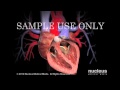 Aortic Valve Replacement
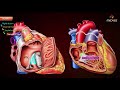 Gross anatomy of Right atrium (RA) - Medvizz Anatomy animated medical videos Usmle step 1
 EKG Interpretation - Master Basics Concepts of ECG
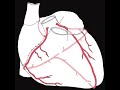 Coronary circulation of the heart
 Abdominal organs (plastic anatomy)
 You and Your Stent
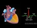 Cardiac Conduction System and Understanding ECG, Animation.
 Cardiovascular System 1, Heart, Structure and Function
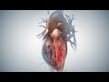 Acute Coronary Syndrome and Heart Attack
 Cardiovascular | Structures and Layers of the Heart
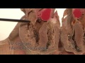 Heart and Pericardium
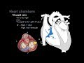 Overview of Heart Anatomy Tutorial
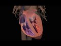 Heart Failure
 EKG/ECG Interpretation (Basic) : Easy and Simple!
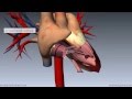 Heart Anatomy - Right Ventricle - 3D Anatomy Tutorial
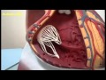 External and internal features of the heart - plastic model
 Anatomy of the Heart: Ventricles, Atria and Functions - Human Anatomy | Kenhub
 Human Circulatory System
 Anatomy of the Heart