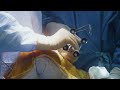
Arthroscopic Transosseous Repair of Medial Meniscal Posterior-Root Tear: Pass Sutures
Video 7 of 10 from the JBJS EST article, Arthroscopic Transosseous Repair of a Medial Meniscal Posterior-Root Tear, by Nicholas P. Gannon, Kelsey L. Wise, Jeffrey A. Macalena. Published October 7, 2021.
➡️ https://bit.ly/3zmV922
Journal: JBJS Essential Surgical Techniques
➡️ https://jbjs.org/journal.php?j=est
Subspecialties: Knee, Sports Medicine
Background: Meniscal root tears are soft-tissue and/or osseous injuries characterized by an avulsion of, or tear within 1 cm of, the native meniscal insertion1. These injuries account for 10% to 21% of all meniscal tears, affecting nearly 100,000 patients annually. Medial meniscal posterior-root tears (MMPRTs) expose the tibiofemoral joint to supraphysiologic contact pressure, decreased contact area, and altered knee kinematics similar to a total meniscectomy3. This injury predisposes the patient to exceedingly high rates of osteoarthritis and total knee arthroplasty secondary to an inability to resist hoop stress4. The arthroscopic transosseous repair of an MMPRT is described in the present article.
Description: (1) Preoperative evaluation, including patient history, examination, and imaging (i.e., radiographs and magnetic resonance imaging). (2) Preparation and positioning. The patient is placed in the supine position, and anteromedial and anterolateral portals are created. (3) Placement of sutures. Two simple cinch sutures are placed into the posterior horn, within approximately 5 mm of each other. (4) Footprint decortication. Remove articular cartilage from the native root insertion site. (5) Drilling of the transosseous tibial tunnel. Introduce a tibial tunnel guide over the decorticated base, set guide to 45° to 50°, place a 2-cm vertical incision over an anteromedial tibial guide footprint, advance a 2.4-mm guide pin through the guide, and overream to 5 mm. (6) Passing of the sutures with use of a looped suture passer introduced retrograde through the tibial tunnel to retrieve sutures. (7) Anchor placement and fixation. Apply maximum suture traction, drill a second aperture 0.5 to 1.0 cm distal to the original aperture on the anteromedial aspect of the tibia, pass the suture ends through the anchor, and fix the anchor into the aperture. (8) Repair evaluation and closure. Note the position and stability of the meniscal root relative to the native footprint. Standard closure in layers is performed.
Keywords: JBJS, EST, Essential Surgical Techniques, Key Procedures, medial meniscus posterior root tears, MMPRT, indirect suture anchor repair, indirect transosseous transtibial repair, pin placement, overreaming, reamer, straight suture passer, stiffened suture, tibial tunnel, suture retriever, soft tissue bridge, cannula, meniscus, tibia
Видео Arthroscopic Transosseous Repair of Medial Meniscal Posterior-Root Tear: Pass Sutures канала JBJSmedia
Показать
Комментарии отсутствуют
Информация о видео
Другие видео канала
 Robotic-Arm-Assisted Lateral Unicompartmental Knee Arthroplasty with a Fixed-Bearing Implant
 Orthopaedic Trauma Research Priorities in Latin America
 Properties of Grafts Commonly Used for Cruciate Ligament Reconstruction
 OrthoJOE Podcast (Episode 6) How is Data Redefining Surgery?
 High-Volume Arthroplasty Centers Demonstrate Higher Composite Quality Scores and Enhanced Value
 Rotator Cuff Degeneration
 59. Leadership, Changes in Shoulder Surgery, and JBJS Open Access, with Special Guest Robin Richards
 Revision Fw Cemented & Uncemented Oxford-III Primary Medial Unicompartmental Knee Replacements
 Episode 44. Hot Topics in Orthopaedics: Oxygen and Ozone
 Comparison of “Human” and AI Hand-and-Wrist Skeletal Age Estimation in Epiphysiodesis
 28. Brandt and the Dude on Urinary Catheters and PROMS in Total Joints
 Dr. Marc Swiontkowski on choosing where to submit a manuscript
 JBJS Miller Review Course
 Number of Levels of Spinal Fusion Associated with the Rate of Hip Joint-Space Narrowing
 JBJS Clinical Classroom on NEJM Knowledge+
 JBJS JOPA Tutorial
 Association Between Knee Alignment and Meniscal Tear in Pediatric Patients with ACL Injury
 Episode 36. AAOS Clinical Practice Guidelines on OA of the Knee (3rd Edition)
 JBJS.org: My JBJS Personalization
 Episode 43. Physician Etiquette and Attire (with Special Guests Elizabeth Arendt and Herman Johal)