Fetal echocardiography at 11-13 weeks: Coarctation of the Aorta (CoA)
www.fetalechocardiography.com
This video clip describes echocardiographic features of coarctation of the aorta (CoA) in fetus at 12 and 13 weeks of pregnancy. The fetus had Turner syndrome. There is also persistent left superior vena cava (PLSVC), which can be associated with CoA.
Видео Fetal echocardiography at 11-13 weeks: Coarctation of the Aorta (CoA) канала Fetal Echocardiography
Показать
Комментарии отсутствуют
Информация о видео
Другие видео канала
 Fetal echocardiography at 11-13 weeks: Ventricular Septal Defect (VSD)
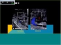 Topics in Fetal Medicine: Left-sided Obstructive Lesions in the Fetal Heart
 Fetal echocardiography at 11-13 weeks: Tetralogy of Fallot (TOF)
 Aortic coarctation (adult) ECHO FINDINGS
 Fetal echocardiography at 11-13 weeks: Right Aortic Arch (RAA)
 Abdominal Aortic Aneurysm (AAA) Ultrasound
 Fetal echocardiography at 11-13 weeks: Ectopia Cordis in Pentalogy of Cantrell
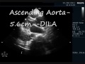 DISSECTION OF AORTA - ECHOCARDIOGRAPHY SERIES BY DR ANKUR K CHAUDHARI.
 Adult Congenital Heart Disease: Anomalous Pulmonary Veins
 Atrial Septal Defect: X-ray, ECG, Echo, TEE, Device Closure
 Fetal Echocardiography Masterclass 2022
 Atrioventricular Septal Defects (Ami Bhatt, MD)
 What the Echo Doesn't See | Echo Challenges
 Basics in Perinatal Neurosonography
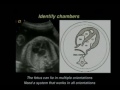 Efficient and Effective Interpretation of the Four Chamber Heart View and Views of the Great Arterie
 CCUS: Pulmonary Hypertension Assessment
 Fetal echocardiography at 11-13 weeks: Pericardial Effusion
 Ultrasound Video showing soft tissue edema of the fetal body.
 Ultrasound of Aortic Dissection
 Echocardiography of Coarctation of the Aorta & Interrupted Aortic Arch