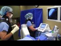
Dr. McRoberts performs a Facet Joint Nerve Ablation in Real Time With a Real Patient.
I have many patients ask me what it is like to have a facet joint nerve ablation or "rhizotomy" and so I decided to video one.
Herein is a video of a patient enjoying a "FJNA."
The process is fairly simple. In most people the anatomy of the facet joint nerve, also called the "medial branch of the dorsal primary ramus" in the lumbar and cervical spine is in the same position, and so its easy to find the location on X-ray or fluoroscopy.
We identify the nerves which innervate the painful joints and then pass a thin needle down to the nerve. The tip of the needle gets hot as we increase the temperature using very precise radio frequency. It forms a spindle shaped lesion about the size of a grain of rice (half a grain in the cervical spine). That lesion interrupts the pain signal in the nerve so that the pain in the joint is no longer felt by the nerve.
This lasts on average around 400 days, sometimes less, sometimes more, then the nerve re-grows back and we have to do the procedure again if need be. Sometimes the arthritic joint has fused by that time and there is no pain. Sometimes the joint arthritis or synovitis has gotten markedly better. Sometimes the pain is the same and we have to re-do the joint.
Regardless, in successful patients the results can be profound with resolution of back and neck pain.
get more info at www.internationalhouseofpain.com
Видео Dr. McRoberts performs a Facet Joint Nerve Ablation in Real Time With a Real Patient. канала Anodyne Research
Показать
Комментарии отсутствуют
Информация о видео
Другие видео канала
 Third Occipital Nerve Radiofrequency Ablation
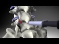 Facet Injection and Medial Branch Nerve Block Patient
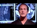 Epidural Steroid For Lumbar Spinal Stenosis Pros and Cons by Dr. Tony Mork
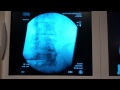 Radio Frequency Ablation Demonstration
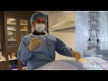 Spinal Cord Stimulator Trial
 Top 3 Signs Your Back Pain is Facet Joint Syndrome-Symptoms & Signs
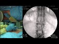 Live Demonstration – Lumbar Spinal Cord Stimulation Trial Brett Stacey, M.D.
 Liver metastases MWA ablation with Emprint™ Ablation System
 Genicular Nerve Ablation for knee pain
 Cervical Facet Radiofrequency Neurotomy - Neck
 Is Facet Joint Syndrome Causing Your Back Pain? EVERYTHING YOU SHOULD KNOW
 Lumbar radiofrequency ablation
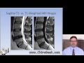 Dr. Gillard lectures on How to Read Your Lumbar MRI
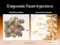 Lumbar Facet Joint Nerve Injections for Treating Chronic Low Back Pain
 Watch Dr. Lynch Perform a Radio Frequency Ablation - Live
 Radiofrequency Ablation for Lower Back Pain, Knee Pain and Headaches
 Dr. Lynch Genicular Nerve Ablation
 Basics of Facet Syndrome (Facet Arthritis) from an AZ pain clinic (602) 507-6550
 Lumbar Spinal Stenosis, Cauda Equina Syndrome, Sciatica, & Disc Herniation: An Advanced Lecture.
 Facet Injection - Dr. Sara Baird