17. Measurement of acceleration time (for mPAP) at right ventricular outflow tract (RVOT)
From the National Pulmonary Hypertension Service Pulmonary Hypertension Echocardiography protocol.
For interactive pdf with embedded clips visit:
www.ph-echocardiography-protocol.com
(Voiceover from David Dawson, Chief Echocardiographer, Hammersmith Hospital, Imperial College Healthcare NHS Trust, London, UK)
Видео 17. Measurement of acceleration time (for mPAP) at right ventricular outflow tract (RVOT) канала Luke Howard
Показать
Комментарии отсутствуют
Информация о видео
Другие видео канала
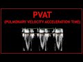 Pulmonary Velocity Acceleration Time (PVAT)! - Echocardiography
 16B. Using pulmonary regurgitation to measure mean PAP and PA end-diastolic pressure
 Critical Care Echo of the RV (Part 3) - Beyond TR jets: Measuring pulmonary hemodynamics
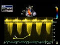 14. Estimating right ventricular / pulmonary artery systolic pressure from TR jet
 MAPSE and TAPSE
 visual estimating LV Ejection Fraction
 25. Measurement of pulmonary vascular resistance
 20. Normal septal motion in systole (LVEI)
 How to...Measure Pulmonary Acceleration Time
 Echo Challenge: A Differential Diagnosis
 60/60 Sign for Acute PE
 23. Myocardial performance index (Tei index) using tissue doppler
 20. Isolated volume loading of the right ventricle (LVEI)
 Top 10 Facts About Right Ventricular Function
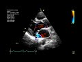 Echocardiographic assessment of the mitral valve
 Pulmonary Imaging: Techniques for Pulmonary Hypertension Diagnosis (Zeenat Safdar, MD)
 Estimate the Right Atrial Pressure using the IVC. Perioperative & Critical Care ECHO/ POCUS
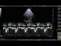 Pulse Wave Doppler Step by Step - LVOT VTI Example
 Residency | Pulmonary Hypertension | @OnlineMedEd
 How to estimate pulmonary artery systolic pressure (PASP) using echo