Cardiac Tamponade: Pathophysiology, Etiology, Symptoms, Diagnosis and Management, Animation
(USMLE topics) Cardiac tamponade: Pathophysiology, causes, symptoms, diagnosis and treatments. This video is available for instant download licensing here: https://www.alilamedicalmedia.com/-/galleries/narrated-videos-by-topics/cardiac-pathology/-/medias/fd49d72a-b1f9-4c54-aeaa-36d54ed199d6-cardiac-tamponade-narrated-animation
Voice by: Ashley Fleming
©Alila Medical Media. All rights reserved.
Support us on Patreon and get early access to videos and free image downloads: patreon.com/AlilaMedicalMedia
All images/videos by Alila Medical Media are for information purposes ONLY and are NOT intended to replace professional medical advice, diagnosis or treatment. Always seek the advice of a qualified healthcare provider with any questions you may have regarding a medical condition.
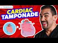
The heart is enclosed in a double-walled, fluid-filled sac called the pericardium. Cardiac tamponade happens when fluid accumulates abnormally in the pericardial sac, to such an extent that it compresses the heart, leading to decreased cardiac output and circulatory shock.
Tamponade may develop from a hemorrhage, such as from a traumatic injury to the chest, ventricular wall rupture after a heart attack, ruptured aortic aneurysm, or a complication after heart surgery. Fluid may also accumulate as a result of other health conditions, such as infections, autoimmune diseases, kidney failure, inflammatory diseases, and cancers.
The rate at which fluid is building up is often more critical than the volume of fluid. Slow accumulation of a large volume may not cause tamponade, but a relatively small effusion can do so if it builds up too rapidly, because the pericardium cannot stretch quickly enough to accommodate it. Traumatic events usually lead to faster fluid accumulation and are therefore more dangerous.
Cardiac tamponade symptoms are typical of a cardiogenic shock, and include: chest pain, rapid heart rates, shortness of breath, and in severe cases: dizziness, fainting, and altered mental status. The skin may feel cold, sweaty, and bluish. Neck veins may appear swollen.
Diagnosis is based on clinical evaluation, typically the presence of Beck's triad: hypotension, muffled heart tones and distended neck veins. Among imaging techniques, bedside echocardiography is the preferred method to visualize and determine the size of effusion.
Tamponade is a medical emergency. Pericardiocentesis is usually the first procedure performed to remove excess fluid from the pericardial cavity. A needle or a small catheter is used to draw the fluid out, preferably under echocardiography guidance. Removal of a small amount of fluid is usually sufficient to stabilize blood pressure, but a catheter can be left within the pericardium to allow for further drainage. Aspiration of fluid, however, may not work for traumatic pericardial effusions, which often consist of blood clots. Surgical options including creating a pericardial window or removal of the pericardium, are preferred in this situation.
Видео Cardiac Tamponade: Pathophysiology, Etiology, Symptoms, Diagnosis and Management, Animation канала Alila Medical Media
Voice by: Ashley Fleming
©Alila Medical Media. All rights reserved.
Support us on Patreon and get early access to videos and free image downloads: patreon.com/AlilaMedicalMedia
All images/videos by Alila Medical Media are for information purposes ONLY and are NOT intended to replace professional medical advice, diagnosis or treatment. Always seek the advice of a qualified healthcare provider with any questions you may have regarding a medical condition.
The heart is enclosed in a double-walled, fluid-filled sac called the pericardium. Cardiac tamponade happens when fluid accumulates abnormally in the pericardial sac, to such an extent that it compresses the heart, leading to decreased cardiac output and circulatory shock.
Tamponade may develop from a hemorrhage, such as from a traumatic injury to the chest, ventricular wall rupture after a heart attack, ruptured aortic aneurysm, or a complication after heart surgery. Fluid may also accumulate as a result of other health conditions, such as infections, autoimmune diseases, kidney failure, inflammatory diseases, and cancers.
The rate at which fluid is building up is often more critical than the volume of fluid. Slow accumulation of a large volume may not cause tamponade, but a relatively small effusion can do so if it builds up too rapidly, because the pericardium cannot stretch quickly enough to accommodate it. Traumatic events usually lead to faster fluid accumulation and are therefore more dangerous.
Cardiac tamponade symptoms are typical of a cardiogenic shock, and include: chest pain, rapid heart rates, shortness of breath, and in severe cases: dizziness, fainting, and altered mental status. The skin may feel cold, sweaty, and bluish. Neck veins may appear swollen.
Diagnosis is based on clinical evaluation, typically the presence of Beck's triad: hypotension, muffled heart tones and distended neck veins. Among imaging techniques, bedside echocardiography is the preferred method to visualize and determine the size of effusion.
Tamponade is a medical emergency. Pericardiocentesis is usually the first procedure performed to remove excess fluid from the pericardial cavity. A needle or a small catheter is used to draw the fluid out, preferably under echocardiography guidance. Removal of a small amount of fluid is usually sufficient to stabilize blood pressure, but a catheter can be left within the pericardium to allow for further drainage. Aspiration of fluid, however, may not work for traumatic pericardial effusions, which often consist of blood clots. Surgical options including creating a pericardial window or removal of the pericardium, are preferred in this situation.
Видео Cardiac Tamponade: Pathophysiology, Etiology, Symptoms, Diagnosis and Management, Animation канала Alila Medical Media
Показать
Комментарии отсутствуют
Информация о видео
Другие видео канала
 Cardiac Tamponade NCLEX Tips for Nursing Students
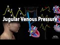 Jugular Venous Pressure, Animation
 PERICARDIAL EFFUSION vs CARDIAC TAMPONADE - EXPLAINED IN 5 MINUTES (Beck's Triad, Causes, Diagnosis)
 Tension Pneumothorax – Emergency Medicine | Lecturio
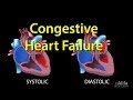 Congestive Heart Failure: Left-sided vs Right-sided, Systolic vs Diastolic, Animation.
 Acute Coronary Syndrome: Unstable Angina, NSTEMI and STEMI (Heart Attack), Animation
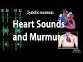 Heart Sounds and Heart Murmurs, Animation.
 Chest Trauma: Cardiac Tamponade
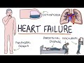 Understanding Heart Failure: Visual Explanation for Students
 The Cardiac Cycle, Animation
 Pericarditis/ Tamponade physical findings
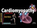 Cardiomyopathy, animation
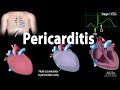 Pericarditis: Symptoms, Pathophysiology, Causes, Diagnosis and Treatments, Animation
 Echocardiogram of a Patient With Cardiac Tamponade
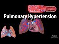 Pulmonary Hypertension, Animation
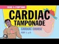 Cardiac Tamponade: Pathophysiology & Symptoms for Nursing Students & NCLEX (1 of 5)
 Cardiac Tamponade- Pulsus Paradoxus
 Emergency Pericardial Centesis Procedure Explained
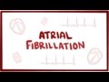 Atrial fibrillation (A-fib, AF) - causes, symptoms, treatment & pathology
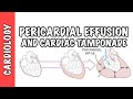 Cardiac Tamponade - pericardial effusion, causes, pathophysiology, investigations and treatment