Acute Otitis Media: Otoscopic Findings
Acute otitis media can take on a variety of appearances with a range of different colors. Otoscopic findings include alterations in translucency, position, and mobility; and other features such as air bullae, fluid levels, and perforations. Erythema of the tympanic membrane is a common finding in patients with acute otitis media, however it is NOT by itself diagnostic of an infection. Patients with a cough or a fever may also be noted to have a reddened eardrum. Appreciation of the limitation of this finding is important in the prevention of inappropriate antibiotic prescription. This second example features a bulging tympanic membrane, which is the hallmark of acute otitis media. It is the single most specific sign of an infection of the middle ear. This third tympanic membrane is bulging, opaque, and covered by a thing layer of discharge. This combination of findings is highly predictive of acute otitis media. Opacity or cloudiness can also be seen in otitis media with effusion and cannot in isolation be used to distinguish between the two conditions. The fourth example features air bullae, which is indicative of bullous myringitis, an ear infection that can be exquisitely painful. In addition to these findings, immobility detected by pneumatic otoscopy can provide evidence of a middle ear effusion. This can be particularly helpful in cases where the tympanic membrane is in a neutral position and yet appears to be inflamed.
0:00 Intro
0:17 Erythema
0:36 Bulging tympanic membrane
0:47 Bulging and opaque tympanic membrane
1:04 Air bullae
🌐 Check out our website for more video lectures
https://www.med4vl.com
📺 Subscribe To My Channel and Get More Great Quizzes and Tutorials
https://www.youtube.com/channel/UC95TzSH1B_2EjaZMgDBNmvA?sub_confirmation=1
Disclaimer: All the information provided by Medical Education for Visual Learners and associated videos are strictly for informational purposes only. It is not intended as a substitute for medical advice from your health care provider or physician. It should not be used to overrule the advice of a qualified healthcare provider, nor to provide advice for emergency medical treatment. If you think that you or someone that you know may be suffering from a medical condition, then please consult your physician or seek immediate medical attention.
Видео Acute Otitis Media: Otoscopic Findings канала Medical Education for Visual Learners
Показать
Комментарии отсутствуют
Информация о видео
15 августа 2018 г. 14:01:04
00:01:46
Другие видео канала
 Acute Otitis Media (Causes, Pathophysiology, signs and symptoms, treatment and complications)
 Tympanic Membrane Anatomy - Head and neck , Medvizz Anatomy medical animations
 Weber and Rinne Test - Clinical Examination
 OTOSCOPY OSCEs | PLAB Image Reference
 Ruptured Eardrum | Tympanic Membrane Perforations
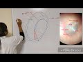 Normal Tympanic Membrane
 Otoscopic Signs of Acute Otitis Media
 Otitis Media: Anatomy, Pathophysiology, Risk Factors, Types of OM, Symptoms and Treatment, Animation
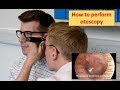 How to perform Otoscopy (Ear Exam)
 Otoscopy and Hearing Assessment | Ear Examination | Rinne’s & Weber's test | OSCE Guide
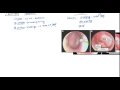 Acute Otitis Media and Otitis Media with Effusion for USMLE Step 2
 Chest X-ray Quiz: Low Difficulty
 Middle Ear Infection (Acute Otitis Media) | Causes, Symptoms, Diagnosis, Treatment
 Otoscopy (Ear Examination) - ENT
 Special Senses | External & Middle Ear Anatomy
 ECG Quiz: Medium Difficulty
 Acute Otitis Media: Otoscopy
 Brain CT Quiz: Medium Difficulty
 Ear Pain 5: Otoscope Examination
 ECG Quiz: High Difficulty