EVALUATION OF MITRAL VALVE AT TRANSESOPHAGEAL ECHOCARDIOGRAPHY (TEE)
When we evaluate the mitral valve at the transesophageal echocardiogram (TEE) we should evaluate mitral anatomy, the leaflets and the annulus in multiple views, so we have to scan the valve through multiple angles.
We start at zero degrees in four-chambers view, where we can see, from the left-hand side to the right-hand side, A3, A2, P2 and P1.
Then we continue at 60-degree in the bicommissural view, in which we can see P3, A2 and P1, where P3 is medial, close to the coronary sinus and P1 is lateral, close to the appendage.
Then, the 90-degree view, in which we see P3, A2 and A1.
Finally we look at the 120-degree view with P2 and A2, together with the aortic valve.
After that, we evaluate all the previous views with color to evaluate the jet and the direction of the mitral regurgitation.
Moreover, we can also check for the 3D vena contracta area.
Finally, we look at the 3D surgical mitral valve view. We have the aortic valve at 12-o’clock. On the left we have the left appendage, which is lateral. On the right we have the septum and tricuspid valve which are medial. The anterior and posterior leaflet could be divided into three scallops. From the left to the right we have A1, A2 and A3 for the anterior leaflet as well as P1, P2 and P3 for the posterior leaflet. Hence, A1 and P1 are lateral, A3 and P3 are medial.
Please note: All echocardiographic images or videos are anonymous, and when a single case is presented written consensus of the patient has been collected for the publication of the images.
Видео EVALUATION OF MITRAL VALVE AT TRANSESOPHAGEAL ECHOCARDIOGRAPHY (TEE) канала Sergio Suma
Показать
Комментарии отсутствуют
Информация о видео
Другие видео канала
 How to optimize a 3D acquisition at transesophageal echocardiography
 Mitral valve surgery - Dr Suad El Qarra
 Contrast and stress echocardiography - Dr Nicola Gaibazzi
 WHAT IS GLOBAL LONGITUDINAL STRAIN AND ITS CLINICAL IMPORTANCE?
 MITRAL REGURGITATION TRANSESOPHAGEAL ECHOCARDIOGRAPHY (TEE) - CASE 5
 MITRAL REGURGITATION TRANSESOPHAGEAL ECHOCARDIOGRAPHY (TEE) - CASE 4
 MITRAL REGURGITATION TRANSESOPHAGEAL ECHOCARDIOGRAPHY (TEE) - CASE 3
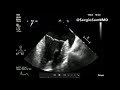 MITRAL REGURGITATION TRANSESOPHAGEAL ECHOCARDIOGRAPHY (TEE) - CASE 2
 MITRAL REGURGITATION TRANSESOPHAGEAL ECHOCARDIOGRAPHY (TEE) - CASE 1
 EVALUATION OF GLOBAL LONGITUDINAL STRAIN (GLS) AT THE TRANSTHORACIC ECHOCARDIOGRAM (TTE)
 EVALUATION OF 3D LEFT VENTRICULAR VOLUMES AT TRANSTHORACIC ECHOCARDIOGRAPHY (TTE)