Sacroiliac Joint Dysfunction Anatomy, Animation - Everything You Need To Know - Dr. Nabil Ebraheim
Dr. Ebraheim’s educational animated video describes the anatomy of the sacroiliac joint.
The sacroiliac joint lies between the sacrum and the ilium. The sacrum lies at the lower part of the spine. The SI joint may be a source of low back pain, however any structure in the lower back may cause pain such as:
•Facets
•Intervertebral discs
•Spinal canal
•Muscles
•Ligaments
All forces from the lumbar spine are transmitted to the sacrum. These forces then move sideways into the SI joints, the pelvis, the hips and the lower extremities. The sacrum is connected to the pelvis by ligaments. The interosseous sacroiliac ligaments are strong ligaments seen dorsal to the cavity of the SI joint at its narrow recess between the sacrum and the ilium. The posterior sacroiliac ligaments are located behind the interosseous sacroiliac ligaments. The anterior sacroiliac ligament covers the anterior portion of the SI joint.
The other posterior ligaments:
1-Sacrospinous ligament: from the sacrum to the ischial spine. Gives rotational stability.
2-Sacrotuberous ligament: from the sacrum to the ischial tuberosity. Gives vertical stability. Both the sacrospinous and sacrotuberous ligaments anchor the sacrum to the ischium.
3-Iliolumbar ligament runs from the transverse process of L5 to the anterior superior iliac crest close to the PSIS. Avulsion fracture of the transverse process of L5 on x-ray is a clue for pelvic injury.
The sacrum has a thick articular surface covered in smooth hyaline cartilage. The articular surface of the sacrum is an ear shaped irregular surface with ridges, prominence, and depressions. The articular surface of the sacrum is matched to the articular cartilage of the ilium, which is made up of thin cartilage. The SI joint is near an important site for bone graft harvesting. Anteriorly to the SI joint, the L4-L5 nerve roots pass close to the SI joint. The pelvis is a ring and the sacrum is a part of that ring.
Movement of the SI joint
The SI joint is a complex structure composed of two distinct compartments. The posterior compartment is ligamentous, and the anterior compartment is synovial. Authors and clinicians disagree about the function, movement and the problems of the SI joint. However, everyone agrees that stability and strength of the joint are important to transmit the force. The SI joint is a bony interlocking mechanism with strong ligaments absorbing all the twisting movements. Some people believe that the SI joint moves by stretching the ligaments and that this movement decreases with age (joint becomes stiff). The joint slides or translates about 2 mm and rotates about 3 mm. excessive movement is abnormal. The SI joint can be a source of pain and dysfunction.
Differentiation between SI joint pain and other sources of low back pain may be difficult.
The history and clinical exam alone is suggestive. Faber test may even be unreliable. The only confirmatory test for problems of the SI joint is with relief of pain after intra-articular injections.
Become a friend on facebook:
http://www.facebook.com/drebraheim
Follow me on twitter:
https://twitter.com/#!/DrEbraheim_UTMC
Видео Sacroiliac Joint Dysfunction Anatomy, Animation - Everything You Need To Know - Dr. Nabil Ebraheim канала nabil ebraheim
Показать
Комментарии отсутствуют
Информация о видео
Другие видео канала
 Simple Solutions to Sacroiliac (SI) Joint Pain
 I have SIJ Dysfunction- MRI is normal and Physical Therapy didn't help- Is Prolotherapy an option?
 Sacroiliac Joint Movement: Nutation II Counternutation II Sacral Torsion II Iliac Torsion
 Pelvic Fractures - Everything You Need To Know - Dr. Nabil Ebraheim
![The Sacroiliac Joint [Part 1] | Major Ligaments & Structures](https://i.ytimg.com/vi/fNpIEzhEt1A/default.jpg) The Sacroiliac Joint [Part 1] | Major Ligaments & Structures
The Sacroiliac Joint [Part 1] | Major Ligaments & Structures SI Joint Diagnosis - History and Patient Presentation - John Swofford, DO
 Diagnosis and Treatment of the Sacroiliac Joint - Charles Harvey, MD
 3 Exercises for SI Joint Pain Relief
 Piriformis Syndrome A Hidden Cause of Sciatica - Everything You Need To Know - Dr. Nabil Ebraheim
 Posterior Pelvic Ring Dr Conor Kleweno
 Understanding Sacroiliac Joint Pain
 Vagus nerve Anatomy Animation / Cranial nerve X : Origin, Course, Nuclei, Branches - Neuroanatomy
 Coronoid Process Fracture Treatment ,approaches Everything You Need To Know - Dr. Nabil Ebraheim
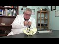 Ligament Healing for Chronic Sacroiliac Joint Dysfunction SIJD
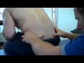 How to assess motion of the Sacroiliac Joint - Seated forward flexion test
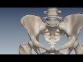 SI Joint Anatomy, Biomechanics & Prevalence
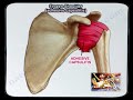 Frozen Shoulder Adhesive Capsulitis - Everything You Need To Know - Dr. Nabil Ebraheim
 Chronic SI (Sacroiliac) Joint Pain? Your Feet May Be The Problem.
 Do you have a Sacroiliac Joint Problem? Learn about how we test for Sacroiliac joint (SI) problems
 Sacroiliac Joint Pain: Diagnosis and Treatments