MRI Flow and Function
Lecture about performing cardiac flow and function on MRI.
Table of Contents:
00:11 - Financial Disclosure
00:21 - k-space
00:26 -
01:02 - How often do you do flow and functional analysis?
01:19 - Image Acquisition processing
01:24 - Phase contrast imaging
02:10 - Do you use a calibration phantom?
02:37 - gating
03:34 - Breath-hold vs free breathing
04:08 - Which technique do you most commonly use for flow imaging?
04:19 - Velocity Encoding
05:37 - Velocity encoding
06:25 - In-plane flow
06:41 - Setup through-plane flow image
06:49 - Through-plane flow
06:58 - Measuring flow
07:41 - Tips
08:21 - volume and mass
09:09 - Auto or semi-auto segmentation
10:02 - Full functional analysis
11:47 - Myocardial Mass
12:44 - Warning
12:49 - Function formulas
13:13 - Clinical Applications
13:18 - Clinical applications
13:52 - Stenosis with gradient
13:54 - Performing flow analysis
14:47 - Aortic Stenosis
15:18 - Aortic Stenosis
15:47 - What is grade of the aortic stenosis?
16:02 - Stenosis classification
16:55 - Aortic coarctation
17:05 - Aortic coarctation
18:15 - Regurgitant valves
18:19 - Pulmonic Regurgitation
18:49 - Pulmonic Regurgitation
19:17 - What is the grade of the regurgitation?
19:27 - RegurGitant valve classification
19:41 - Mitral regurgitation
20:09 - What 2 values will allow calculation of the mitral regurgitant fraction?
20:37 - Mitral regurgitation
21:24 - cardiomyopathy
21:26 - Septal hypertrophy
21:53 - Subvalvular flow
21:57 - What is the gradient across the subvalvular stenosis?
22:34 - Congenital Shunt
22:39 - Congenital with shunt
22:49 - Congenital with shunt
23:20 - Learning objectives "It's like deja vu all over again." -Yogi Berra
23:39 - Thank You
Видео MRI Flow and Function канала Shawn Teague
Показать
Комментарии отсутствуют
Информация о видео
Другие видео канала
 Cine MRI for Chiari I Malformation
 MRI Imaging Planes
 MRI: Basic Physics & a Brief History
 Are MRIs safe?
 Diseases of the Aorta: A Multimodality Approach/CT and MRI (Dr. Quinones, MD/Dr. Nabi, MD) 02/21/17
 How to assess aortic stenosis using CMR
 The Role of Cardiac MRI in Heart Failure (Dipan J. Shah, MD)
 Introducing MRI: Phase Contrast MRA (45 of 56)
 Cardiac Magnetic Resonance Imaging (MRI) Basic Principles (Dipan Shah, MD) Sep. 29, 2015
 Introducing MRI: Chemical Shift (28 of 56)
 Cardiac MRI: Basic Concepts and New Developments (JOÃO L. CAVALCANTE, MD)
 The fundamentals of left ventricular assessment in cardiac magnetic resonance imaging (CMR)
 TMT: CSF Flow Studies Part IV: Normal Pressure Hydrocephalus
 Coronary Anomalies
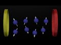 Diffusion Weighted Imaging
 Assessment of RV Function: CMR (ERIC Y. YANG, MD) February 13, 2018
 cvi42 Tips n Tricks 2016
 Susceptibility Weighted Imaging of the Brain - David Mark Yousem
 Stress MRI: Methodology, Current Role and Case Studies (Dipan Shah, MD) December 13, 2016
 Post Processing Cardiac CT