Microscope and its working - Science
This video Microscope and its working is a science lesson/ tutorial for Grade 8-10 students.
Microscope
Microscope is an optical device which makes image of small object to appear very large.
The image is said to be magnified when the size of the image is larger than the size of the object.
The image is said to be magnified when the ratio size of the image is larger than the size of the object.
The ratio of the size of the image represented as v to the size of the object represented as u is called magnification factor.
i.e.
v/u = M
Similarly, if the size of the image is smaller than the size of the object, then the image is said to be diminished.
For v/u = M, if M is greater than 1, then image is magnified.
And if M is lesser than 1, then image is diminished.
Look at the word Physics on a white sheet of paper.
Now, take a convex lens and adjust the distance between the word on the board and the lens suitably so that the letters of the words physics appear big.
Hence, a convex lens is also called a simple microscope.
Let us understand the principle and working of a compound microscope.
To obtain a greater magnification, an optical device called compound microscope is used.
A compound microscope is a combination of two convex lens whose focal lengths are different.
The lens with the short focal length (f) will be facing the object.
This lens is called objective.
The lens with large focal length (F) is used for viewing the object.
This lens is called eye piece.
The two lens are so arranged that their optic centres lie on the same line.
The objective in the eye piece are fitted to one end of two separate tubes.
The two tubes are such that one tube fits into the other and slides freely.
Seeing an inverted image is not a disadvantage as the microscope is used to observe very small objects such as bacteria, blood cells, to measure the diameter of a capillary or cross section of a thin wire.
Видео Microscope and its working - Science канала Elearnin
Показать
Комментарии отсутствуют
Информация о видео
Другие видео канала
 How a compound microscope works? / 3D animated
 Microscopy techniques basics | Microscopy lecture | magnification and resolution of a microscope
 Microscope for Beginners - Questions and Answers
 NEET Physics Concepts Explained | Telescope
 How Lenses Function
 2 The Principle of the Electron Microscope
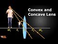 Convex and Concave Lenses
 Microscopes and How to Use a Light Microscope
 How to Choose the Right Microscope Objective
 Fractional Distillation Petroleum Oil Diesel Kerosene
 Properties of Laser - Monochromaticity - Physics
 Transistors, How do they work?
 How Do Microscopes Work? MICROSCOPE Science!
 Using a microscope The parts and how to focus
 Diffraction interference patterns with phasor diagrams
 Fiber optic cables: How they work
 Microscope Parts And Its Functions | Microscope View | Microscope In Hindi | Grow Your Talent
 MICROSCOPE WORKING IN ANIMATION
 Human Eye and Colourful World Class 10