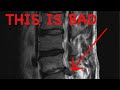
Radiological Anatomy of the Lumbar Spine: X-ray, MRI & CT Covered
In this video, Dr. Gillard lectures on the fundamentals of x-ray (how it works) and then the detailed anatomy of the lumbar spine as shown on X-Ray, MRI and CT imaging. Some common conditions of the lumbar spine will also be briefly covered. This is mandatory material for 8th quarter!
Видео Radiological Anatomy of the Lumbar Spine: X-ray, MRI & CT Covered канала Douglas Gillard, DC, Professor of Clinical Science
Видео Radiological Anatomy of the Lumbar Spine: X-ray, MRI & CT Covered канала Douglas Gillard, DC, Professor of Clinical Science
Показать
Комментарии отсутствуют
Информация о видео
7 августа 2017 г. 9:01:26
01:32:55
Другие видео канала
 How to Read a Spine MRI
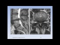 Lumbar Spinal Stenosis, Cauda Equina Syndrome, Sciatica, & Disc Herniation: An Advanced Lecture.
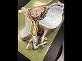 Cervical Spine Anatomy & Some Pathology
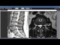 Severe Foot Drop: the Clinical & MRI Findings of a Real Client with a Surprise Finding
 What is Cervical Stenosis? | Jeffrey Cantor, MD
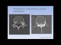 The Spondylolysis / Spondylolisthesis Lecture
 Approach to look at C-spine X-rays
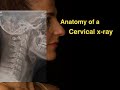 Anatomy of a Cervical x-ray
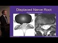 Lumbar MRI: What the Findings Mean and How They Should Be Reported - J. Jarvik, MD, MPH
 Epidural Steroid For Lumbar Spinal Stenosis Pros and Cons by Dr. Tony Mork
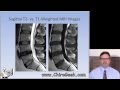 Dr. Gillard lectures on How to Read Your Lumbar MRI
 Adhesive Arachnoiditis: An Advanced Lecture
 Introduction to CT C-spine: Approach and Essentials
 Surgical Procedures - Lumbar Laminectomy & Discectomy
 Anatomy of Knee X-rays
 Anatomy of a Lumbar x-ray
 Management of Cervical Stenosis - Charles A. Sansur, MD
 360°/VR Spine Surgery, Cervical Disc Replacement with Dr. Richard Guyer at Texas Back Institute
 A Case of Double Failed Lumbar Fusion and Dangerously Misplaced Pedicle Screws
 How to Read Your MRI with Onis 2.5 - part 2 of 2 (Advanced Lumbar Spine MRI Anatomy)