Automatic Model-Based Segmentation of Medical Images - Cristian Lorenz Technion lecture
Automatic Model-Based Segmentation of Medical Images
Lecture by Cristian Lorenz of Philips Research Laboratories, Germany
At Technion-Israel Institute of Technology, TCE conference
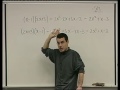
Automatic segmentation of anatomical structures and tracking their changes over time is needed in many medical applications, ranging from cardiology to neurology. Our approach is based on a shape-constrained deformable surface model, implemented as a triangular mesh, that automatically adapts itself to the borders of the subject's anatomy in a 3D medical image. This is done by progressively increasing the degrees-of-freedom of the allowed deformations, improving overall convergence as well as segmentation accuracy. The target anatomy is first localized in the image using the generalized Hough transform. Pose misalignment is corrected by matching the model to the image allowing a global similarity transformation. The initialization of a multi-compartment mesh is then addressed by assigning an affine transformation to each anatomical region of the model. Finally, a deformable adaptation is performed to accurately match the boundaries of the target structure. This presentation describes the underlying methods and gives an overview of clinical applications for various anatomies and imaging modalities like computer tomography (CT), magnetic resonance imaging (MRI) as well as ultrasound (US), each imposing different challenges and requirements to our image processing framework.
Видео Automatic Model-Based Segmentation of Medical Images - Cristian Lorenz Technion lecture канала Technion
Lecture by Cristian Lorenz of Philips Research Laboratories, Germany
At Technion-Israel Institute of Technology, TCE conference
Automatic segmentation of anatomical structures and tracking their changes over time is needed in many medical applications, ranging from cardiology to neurology. Our approach is based on a shape-constrained deformable surface model, implemented as a triangular mesh, that automatically adapts itself to the borders of the subject's anatomy in a 3D medical image. This is done by progressively increasing the degrees-of-freedom of the allowed deformations, improving overall convergence as well as segmentation accuracy. The target anatomy is first localized in the image using the generalized Hough transform. Pose misalignment is corrected by matching the model to the image allowing a global similarity transformation. The initialization of a multi-compartment mesh is then addressed by assigning an affine transformation to each anatomical region of the model. Finally, a deformable adaptation is performed to accurately match the boundaries of the target structure. This presentation describes the underlying methods and gives an overview of clinical applications for various anatomies and imaging modalities like computer tomography (CT), magnetic resonance imaging (MRI) as well as ultrasound (US), each imposing different challenges and requirements to our image processing framework.
Видео Automatic Model-Based Segmentation of Medical Images - Cristian Lorenz Technion lecture канала Technion
Показать
Комментарии отсутствуют
Информация о видео
Другие видео канала
 פולינומים, שורשים ופונקציות רציונליות - 5 - תכונות של חיבור וכפל פולינומים
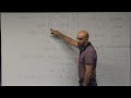 01
01 14
14 24
24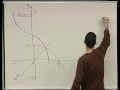 טריגונומטריה - 25 - הפונקציה ארק-קוסינוס
 Swords of Iron – The Robo-physics program
 Rosh Hashanah New Year Experiment from Technion Dipping the Apple in Honey
 Yoav Medan - Ultrasound Surgery - Incision Free Healing - Beyond the Cutting Edge
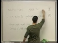 טריגונומטריה - 15 - תכונות של הפונקציה קוסינוס
 Estudiante del Technion International School - Salvador Bentolila - Venezuela
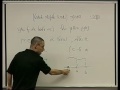 אינטגרלים - 25 - אדיטיביות של האינטגרל המסוים
 Technion International Student Guide - part 3 After Arrival
 Adventures in Public-Key Cryptanalysis - part 1 by Nadia Heninger Technion Lecture
 פולינומים, שורשים ופונקציות רציונליות - 6 - גורמים לינארים
 Rube Goldberg Machine Behind the Scenes Hanukkah 101 Technion Israel
 Technion - Back to the Future, Full Length Version
 וקטורים - 31 - נקודת חיתוך בין ישר למישור
 03
03 The 11th Robotraffic
 חדו"א 2ת' - 44
 05 - Complex numbers