Mastering OCT Interpretation with Dr. Mark Friedberg
In this CEwire2015 highlight, Mark Friedberg, MD gives us an outstanding talk on how to master OCT interpretation.
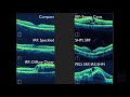
OCT has become an integral tool for many eye care practitioners in the diagnosis and monitoring of retinal disease.
This course will teach how to interpret OCT. Both normal anatomy and pathologic states will be demonstrated.
Finally, numerous mystery cases will be presented to allow the audience to utilize the skills acquired through the course.
Comment here:
https://www.odwire.org/community/threads/mastering-oct-interpretation-with-dr-mark-friedberg.91052/
Видео Mastering OCT Interpretation with Dr. Mark Friedberg канала ODwire.org
Показать
Комментарии отсутствуют
Информация о видео
Другие видео канала
 Macular OCT Interpretation: A Practical Discussion with Dr. David E. Lederer
 Slit Lamp Exam
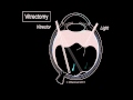 Vitreous 3: Vitrectomy Surgery
 OCT: Interpreting the image
 Visual Field: Practical Guide to Interpretation by Dr. Hannah de Guzman
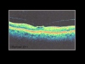 Vitreous 2: Vitreous and Trouble
 How to Read an OCT Image - with Dr. Jerome Sherman
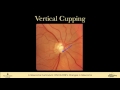 9 EXAMINATION Optic Nerve Head and Nerve Fiber Layer Changes in Glaucoma
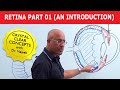 Retina Part 1 - An Introduction
 How to Utilize OCT Angiography to Detect and Manage Diabetic Retinopathy
 The Ophthalmic Exam: Retina and Posterior Segment
 Classification of Diabetic Retinopathy
 Animation: Dilated Eye Exam
 OCT Interpretation Session 3: Variations of Macular Holes
 10 Hacks for OCT Interpretation in Glaucoma - Dr. Mark Dunbar
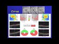 how to read an OCT report of glaucoma ?
 What is Macular Degeneration?
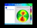 A Quick Guide to Reading Corneal Topography. Part 1
 pitfalls when reading OCT glaucoma report
 OCT Interpretation in the Diagnosis and Management of Glaucoma