cardiac scan with slab misregistration artifact from missing data.
Some CT scanners still require multiple scans at sequential z-axis stations when scanning the heart with prospective triggering and step and shoot technique. During this type of scan, the scanner table has to start and stop its motion multiple times. During acceleration and deceleration, the patient's body may move slightly relative to the table. The patient can also be startled, inducing the patient to move their diaphragm involuntarily. As a result, gaps or redundancy of data may occur in the z-axis dimension. In this case, there is data missing. The heart moved slightly superiorly relative to the table after the first and second scans. Look closely at the midsegment of the LAD for the resulting artifact. This artifact is usually shown with a coronal MPR reconstruction. This is an attempt at 3D visualization. The pink planes are the first and last layers of the slabs at the slab interfaces made nearly opaque after reduction in voxel size after sub voxel sampling.
Видео cardiac scan with slab misregistration artifact from missing data. канала SAVS Volumetric Visualization Laboratory
Видео cardiac scan with slab misregistration artifact from missing data. канала SAVS Volumetric Visualization Laboratory
Показать
Комментарии отсутствуют
Информация о видео
23 декабря 2022 г. 9:43:45
00:00:23
Другие видео канала
 Stenosis ostium of the left main coronary artery and mid segment of LAD
 multifocal disease framesX2 5 speedX1 5
 Air trapping and hepatic steatosis revealed with artificial intelligence noise reduction.
 Cardiac CT AI vs Iterative noise reduction
 important celiac to SMA collaterals
 another common femoral artery
 Metastases and lung anatomy segmented by hand from non contrast scan
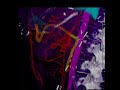 Arc of Riolan in yellow. Occluded celiac and SMA.
 Iliofemoral aa christmas
 prospective scan data
 ribcage framesX2 5 speedX1 5
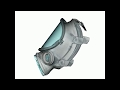 CT of full face respirator from commercial vendor
 aortic insufficiency Interpolated and detail enhanced with AI
 Bullet and streak artifact
 Juxta and infra renal aneurysm
 AI attempt to slow down X10 tachycardia and show tertiary coronary branches #anatomy #coronary
 membranous septum length
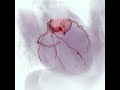 Moderate stenoses diagonal and LAD
 left dominant system, severe hypertension
 First coronary volumetric visualization at UNC Large Ramus