Perimembranous VSD
Please visit my Cardiology Talks channel and subscribe for future updates: https://www.youtube.com/c/JohnsonsCardiologyTalks/
Perimembranous ventricular septal defect is the most common variety. Description at: https://johnsonfrancis.org/professional/echocardiographic-profile-in-ventricular-septal-defect/
Other types are inlet VSD, outlet VSD and muscular VSD. Parasternal long axis view showing right and left ventricles, left atrium and aorta. Perimembranous VSD is seen just below the aortic valve. It is subaortic in location. Size of VSD is compared to the aortic diameter. Here it is a small VSD as it is less than one third of aortic diameter.
Видео Perimembranous VSD канала Johnson Francis, MBBS, MD, DM
Показать
Комментарии отсутствуют
Информация о видео
2 декабря 2020 г. 20:54:50
00:02:24
Другие видео канала
 Supracristal (outlet) ventricular septal defect (V.S.D.) & aortic regurgitation
 VSD-Peri membranus Type. ECHOCARDIOGRAPHY SERIES BY DR ANKUR K. CHAUDHARI
 What is a perimembranous VSD? Intro
 Heart Sounds and Heart Murmurs, Animation.
 ECG Simplified Full Series
 Ventricular Septal Defect, Animation
 Echocardiogram in Ventricular Septal Defect
 "Atrial Septal Defects" by Dr. David Bailly for OPENPediatrics
 Ventricular septal defect (VSD) - repair, causes, symptoms & pathology
 ISUOG 2018 Prof. Julene Carvalho - 'Guidelines Plus' for Fetal Heart Examination
 Perimembranous ventricular septal defect (V.S.D.) Echo (TTE)
 Types of VSD - Hatem Hosny
 ¿infiltrative cardiomyopathy? ¿hypertrophic cardiomyopathy?
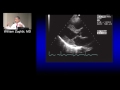 Principles of Echo Doppler (William Zoghbi, MD) September 13, 2016
 ATRIO VENTRICULAR CANAL DEFECTS | MARK SKLANSKY | AVSD | FETAL ECHO | FETAL CARDIAC ANOMALY | DOWN'S
 "Ventricular Septal Defects" by Dr. David Bailly for OPENPediatrics
 MITRAL VALVE PROLAPSE-ECHOCARDIOGRAPHY SERIES BY DR.ANKUR.K.CHAUDHARI
 VSD | DR MARK SKLANSKY | 4 CHAMBER VIEW ANOMALIES | INLET- OUTLET VSD | FETAL ECHO | CARDIAC ANOMALY
 Atrial Septal Defect: X-ray, ECG, Echo, TEE, Device Closure
 Chapter- 11 of 24 Right ventricular outflow tract obstruction