Making sense of your echo report
This video is about Making sense of your echo report
Lets go through the information that the report will contain - When you have an echo you will also have a single lead ECG done at the same time and this is to allow the tallying of mechanical activity with electrical activity so usually the first thing on the echo report will be some mention of the heart rhythm and rate on the ECG during the scan.
Next: The left atrium: this structure is the left atrium and what we have started realising is that the left atrium gives you great insight into your overall heart health. Healthy hearts usually have normal sized left atria. What is normal? About 4 cm across. What does it mean if the left atrium is enlarged…usually it means that the heart is having to work harder than it needs to. Also patients who have AF will have larger left atria.Is it dangerous - no not at all but it does mean that you just need to work out why the heart is working harder and manage accordingly.
The next structure is the Mitral valve. This is this valve here. Now most normal valves are described as being thin and mobile. if the valve is described as being thickened, calcified and restricted then that tells you the valve is structurally abnormal. Either it has been subjected to wear and tear from old age or it has been damaged due to an infection such as rheumatic fever. Functionally valves can still be normal or they can be thickened to the point that they are narrowed and impede blood from passing through in which case they are described as being stenosed or stenosis.
The left ventricle. This is the most important chamber of the heart because it is responsible for pumping blood to the whole of the body and patients who have a weak left ventricle have a much worse prognosis than patients who dont.
So the echo will firstly comment on the size of the left ventricle. Normal size is dependant on body surface area but in general you want the left ventricle at its widest to be no more than 5.5 cm in women and 6 cm across in men. if the ventricle looks bigger than that then you worry that it is being stretched either because of a leaky valve or because it was damaged in some way. The problem is the more it stretches the more likely it is to lose its stretchability in the long run and therefore it can weaken.
So there will be some comment on the echo report on the size of the ventricle. As long as it is not dilated you can feel very comfortable and reassured. The second thing is a mention of something called regional wall motion abnormality. Different parts of the left ventricle receive blood from different blood vessels and therefore if a patient has had a heart attack because one of the blood vessels has blocked, then that part of heart muscle will die and therefore not contract normally whereas the other normal parts will and this is described as a regional wall motion abnormality. So if you have no regional wall motion abnormality then you dont have any substantial damage or scar in your heart.
There will also be a mention of function of the heart… how much blood does it pump to with every beat and this is called the ejection fraction. This is a very crude indicator which is usually visually assessed and the normal value is about 60%. if the heart is not pumping out as much blood as you would expect, it is described as left ventricular impairment. Mild left ventricular impairment is if the ejection fraction is 45-55%, moderate is 35-45% and severe is less than 35%. Now many people get very worried because they may have had 2 echo and they say well last time it was described as 62% and now it is 58% - does this mean that it has gotten worse and the answer is definitely not… the ejection fraction is a very crude measure and variations of 5-10% are completely acceptable.
Finally there will be a mention of the thickness of the walls of the feft ventricle. These should usually be no more than 12mm thick and if they are thicker than that it means that either the heart is working harder than it should (high blood pressure) or there is some form of muscle disease which has caused the heart muscle to be thicker than you’d expect and thickened walls are described as left ventricular hypertrophy. if one wall is thickened it is described as asymmetrical and if all walls are thicked it is described as concentric or symmetrical
Видео Making sense of your echo report канала York Cardiology
Показать
Комментарии отсутствуют
Информация о видео
Другие видео канала
 Making sense of your echo report part 2
 Echocardiogram: An ultrasound for your heart
 Why is my BP all over the place?
 Which Artery is Blocked? An Echo Challenge
 How to tell if your breathlessness is being caused by a heart problem
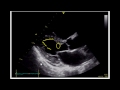 Estimating Ejection Fraction with Point of Care Echo
 HOW TO READ AN ECG!! WITH ANIMATIONS(in 10 mins)!!
 High Blood pressure: Why we shouldnt worry about the number
 Cardiologist discusses reasons why you feel tired and lethargic
 The blood test that detects heart failure
 Slow heart rate or Bradycardia: Will my heart stop?
 Can LVH be reversed?
 3 questions to ask your doctor if he says you have high blood pressure
 The 4 types of heart palpitations
 Palpitations and Beta Blockers
 Heart Disease: Exercise and the heart
 Measuring inflammation - What does CRP tell us?
 Understanding Risk in Afib
 Scared of having a heart attack? Here's what to do about it
 Bedside Ultrasound Basic Cardiac US