Staphylococcal Scalded Skin Syndrome (SSSS) and Bullous Impetigo
Staphyloccocal Scalded Skin Syndrome is caused by Staph infection. Staph bacteria releases two types of toxin Exfoliatin A and Exfoliatin B. Exfolitan A causes more localized infection known as bullous impetigo. Exfolitan B causes the Staphylococcal Scalded Skin Syndrome. It specifically attack desmoglein 1, which is responsible to maintain integrity between spinosum and granulosum.
CLINICALLY there are some specific findings for bullous impetigo is yellow with an erythematous base. This lesion can also be caused by strep pyogenes. Primarily found in exposed areas and orifices. When it ruptures it leaves a red base behind and before rupture they become cloudy vesicle or bulla.
Diagnosis of Bullous Impetigo are from characteritsic lesions however you may aspirate and look for Staphylococcal aureus bacteria.
In Staphylococcal Scalded Skin Syndrome the lesions is profuse and throughout the whole body. Primarily in peri-oral areas and flexures. The lesions are tender and nikolsky's sign positive and the skin would come off. There is a fever present however, they don't look toxic. unless there is a secondary sepsis and pneumonia. Heals within 5-7 days and fast if anti-biotics are given. There is a foci of bacteria residing and it is releasing toxins. It may be in the eye causing purulent conjunctivitis, otitis media, and Nasopharyngeal Infection. Important differential diagnosis such as Steven Johnson Syndrome. but Scalded Skin Syndrome occurs in younger ages. Tzanck Smear so acantholytic lesions and Steven Johnson Syndrome has history of Drug ingestion.
Treatment of Scalded Skin Syndrome is to give emolients and keep skin wet. Fluids do need to be provided as needed (IV).
Eradicate the Staph aureus by giving anti-biotics. Dressings to protect from other infections.
Mortality is around 2% in pediatric population, and 10% in adult population.
Видео Staphylococcal Scalded Skin Syndrome (SSSS) and Bullous Impetigo канала the study spot
Показать
Комментарии отсутствуют
Информация о видео
Другие видео канала
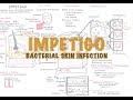 Impetigo Bacterial Skin Infection - Overview (Clinical Presentation, Pathophysiology, Treatment)
 Methicillin Resistant Staphlococcus Aureus Infections
 Steven Johnson Syndrome (SJS), Toxic Epidermal Necrolysis (TEN) and Erythema multiforme for USMLE
 Staphylococcal Scalded Skin Syndrome V.S. Erythema Toxicum Neonatorum
 Staphylococcal Scalded Skin Syndrome – Dermatology | Lecturio
 Acute Respiratory Distress Syndrome (ARDS) for USMLE Step1 and USMLE Step 2
 Staphylococci properties, diseases, diagnosis, and management approach
 Staphylococcal Toxic Shock Syndrome
 Microbiology - Staphylococcus Aureus and Skin Abscess
 IMPETIGO EXPLAINED IN 2 MINUTES - BULLOUS vs NON-BULLOUS IMPETIGO
 "Kawasaki Disease" by Lucy Rubin and Dr. Lisa DiPietro for OPENPediatrics
 Necrotizing Soft Tissue Infections
![Staphylococcal Scalded Skin Syndrome (SSSS) [Med-School Lecture Series]](https://i.ytimg.com/vi/TFfyYTxRPCQ/default.jpg) Staphylococcal Scalded Skin Syndrome (SSSS) [Med-School Lecture Series]
Staphylococcal Scalded Skin Syndrome (SSSS) [Med-School Lecture Series]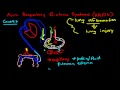 Acute Respiratory Distress Syndrome
 Acute respiratory distress syndrome ( ARDS ) Etiology, Clinical features, Diagnosis, and Treatment
 Serotonin Syndrome vs. Neuroleptic Malignant Syndrome
 2- BACTERIAL INFECTIONS: شرح الجلدية العدوى البكتيريا للجلد د.عبدالرحمن رضا
 The Skin Anatomy, Physiology and Microbiology
 Microbiology - Streptococcus species
 Pulmonary Medicine | Pathophysiology of COPD