Lumbar Spine Pedicle Screw Fixation Fusion lawyer 3D animations
http://www.medilaw.tv Lumbar Spine Pedicle Screw Fixation Fusion lawyer 3D animations. Illustrates the surgical technique for performing a laminectomy and pedicle screw fusion. This procedure is used to decompress the spinal cord and immobilize an intervertebral disc or facet joints that are causing uncontrollable pain. Pedicle screw instrumentation is used to ensure stability while fusion occurs. There are many different techniques to achieve the same end result, a pain-free, stable, anatomically positioned bony fusion. However, the basic procedure illustrated here is common to all pedicle screw fusions. Also shown is the patient position, skin preparation and incision, the surgical approach, the removal of the lamina, the insertion of the pedicle screws, rods and bone graft, x-ray position checks and finally wound closure.
A spinal fusion is done to join two vertebrae together to make one large bone. The surgeon roughens up the external surfaces of the two vertebrae to make the body's natural repair system think that one large bone has broken. The surgeon then adds bone to fill the gap. The body then joins the mass together, like a normal fracture. While the bone is healing, it is held still by screws and plates or rods. Full fusion takes three months. Bone chips can be taken from your hip at the time of the operation, and then grafted onto your vertebra. Alternatively, bone can be harvested from other patients and stored until needed in a bone bank. Using bone from the bone bank saves you the pain of this surgery, but doesn't produce as high fusion rates as using your own bone. Artificial and natural bone substitutes are also available. New bone from the roughened vertebra migrates along the grafted bone to connect the area to be fused. Bone Morphogenetic Proteins may be used to accelerate the fusion rate.
INDICATIONS
A spinal fusion is performed when the spine is unstable, and can't maintain the functional alignment between all of its important structures, or the abnormal movements cause pain and put adjacent structures at risk of injury.
Causes of spinal instability include degenerative joint disease, spondylolysis, fractures, infections and tumors.
Lumbar Spine Pedicle Screw Fixation Fusion lawyer 3D animations
ALTERNATIVES
The non-surgical alternative treatments to lumbar fusion are
avoiding bending, lifting, twisting and prolonged sitting
weight loss
walking
pain-relieving medication
physical therapy
hydrotherapy
The surgical alternative treatments to lumbar fusion are
injections of steroid and local anesthetic around nerves or into the facet joints
lumbar disc replacement in very few cases
The use of lumbar bracing and acupuncture is controversial.
GOALS
The pedicle screw fusion replaces the damaged, painful facet joints with solid bone and the laminectomy gives the compressed spinal cord and nerves more room.
TECHNIQUE
You will be placed in a kneeling position. Your skin will be cleaned. An incision will be made in the middle of the back. The overlying muscles will be moved to the sides. Your surgeon will confirm the correct vertebrae for the procedure by using x-ray imaging. The spinous processes will be removed. The ligamentum flavum is then separated from the lamina. The lamina is removed at each level needing to be decompressed. The remaining ligamentum flavum are removed. The tracks for the pedicle screws are prepared. The vertebra surface is roughened. The bone graft is laid. The remaining pedicle screws, brackets and rods are installed. The muscles are replaced, and the wound is closed with sutures. Lumbar Spine Pedicle Screw Fixation Fusion lawyer 3D animations.
Видео Lumbar Spine Pedicle Screw Fixation Fusion lawyer 3D animations канала medilawtv
Показать
Комментарии отсутствуют
Информация о видео
Другие видео канала
 Anterior Lumbar Interbody Fusion Overview
 Posterior Cervical Laminectomy and Fusion
 Lumbar and Thoracic pedicle screw entry points
 Spine Surgery: What to Expect - Don Park, MD | UCLAMDChat
 STALIF M™ (MIDLINE II) Surgical Animation
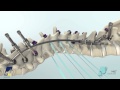 NILE™ Alternative Fixation Spinal System Product Animation
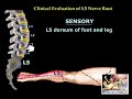 Neurological Evaluation Of The Lumbar Nerve Roots - Everything You Need To Know - Dr. Nabil Ebraheim
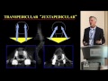 Thoracic Pedicle Screws - Michael Daubs, MD
 Medical Animation of Lumbar Stenosis Decompression
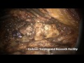 Cervical Pedicle Screw Fixation
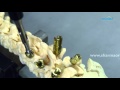 Spondylolisthesis Reduction Screw Fixation- CORE Spinal System
 How to Read a Spine MRI
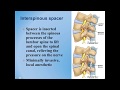 Lumbar Spinal Stenosis : Diagnosis and Treatment Options
 Dorsal and lumber pedicles: surgical anatomy : tips and concerns
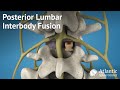 Posterior Lumbar Interbody Fusion Overview
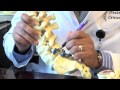 Surgical Procedures - Lumbar Laminectomy & Discectomy
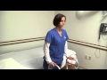 Activity after Spine Surgery
 Medical Minute: BEST Lumbar Spine Broken Hardware Overview, Update
 What is Cervical Stenosis? | Jeffrey Cantor, MD
 Biomechanics of pedicle screw and how they fail