Procedure of Elastography
Every day doctors use ultrasound, CT scan, MRI to see inside of the body to make a decision. Those tests are great that showing size and structure but they don't show physical properties such as tissue stiffness which for example sign of fibroses in a liver. The researcher of Mayo clinic had developed technology that uses sound waves to see if the patients liver is a harder than it should be, if its developing fibroses. It is called magnetic resonance elastography and its offers a non-invasive alternative to liver biopsy.
Magnetic resonans elastography or MRE. It combines MRY technology and low frequency sound waves.
- This is an MRI image of the upper abdomen and here this very large organ in the upper abdomen is the liver. Now, looking at this liver with regular magnetic resonance imaging it is not possible to determine if this patient has fibrosis in the liver.
But when the new MRE technology is used during MRI it shows on the color scale the stiffenes of the liver. Here how its work.
The probe of the Fibroscan device generates the waves in the abdomen when it placed on the patients abdomen. The soundwaves moved through the stiff tissues and normal tissues in at different ways. The computer analyzes the differences and shows what the healthy tissue is and what is not.
This shows the sound waves moving through the tissues of the upper abdomen. And it is show if the patient has liver fibroses or no and where it is localized.
Many liver diseases including hepatitis B, C and other immune hepatitis, alcoholism can lead to fibroses. And if the condition is not successfully treated the disease can progress to cirrhoses for which the only treatment is liver transplantation. MRE is another tool in a fight against the progression of these diseases. In the future the use of MRE may include the imaging of the breast, prostate and other organs.
http://erebouni.am/eng/department/6/Department-of-Ultrasound-Diagnostics
Видео Procedure of Elastography канала Erebouni Medical Center
Показать
Комментарии отсутствуют
Информация о видео
25 января 2016 г. 17:26:20
00:02:14
Другие видео канала
 The FibroScan Technology
 ZP Rad - What Is MRI/PET?
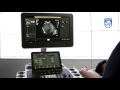 How To Use shear wave Elastography on a Philips EPIQ Ultrasound System
 MR Elastography Webinar
 Liver Elastography Techniques (Part I): RTE & SWM explained
 What to Expect: Magnetic Resonance Enterography (MRE) Scans
 How to eat with cirrhosis of the liver | Ohio State Medical Center
 WHY Sugar is as Bad as Alcohol (Fructose, The Liver Toxin)
 David Sampson: Optical coherence elastography may increase efficiency of cancer surgery
 A patient's introduction to FibroScan | Ohio State Medical Center
 Examination of the jugular venous pulse / JVP examination Procedure video
 How To Perform Strain Elastography on Philips EPIQ Ultrasound Systemsvvv
 GE Healthcare Ultrasound Elastography | GE Healthcare
 IMV imaging Ultrasonography of the Distal Limb video 5 - Basic scanning technique
 "What's new" in Diagnostica per Immagini ELASTOSONOGRAFIA - Introduzione alla Metodica PARTE 2
 Liver Elastography - A Teaching Hospital Experience with Marilyn Zelesco
 What is Attenuation? - www.AcousticFields.com
 Abdominal MRI
 MR Enterography of Inflammatory Bowel Disease with Endoscopic Correlation
 TMT: Elastography by Dr Alka Singhal: Intro to ARFI