Sternoclavicular Joint | Synovial Joint | Type | Attachments | Ligaments | clinical
The sternoclavicular joint is a synovial saddle joint that connects the sternum with the clavicles. It is the only true joint which connects the appendicular skeleton of the upper limb with the axial skeleton of the trunk.
The function of the sternoclavicular joint is to coordinate the movements of the upper limb with the core of the body. Thus allowing the upper limb to perform its full range of motion. Specifically, the movements of the sternoclavicular joint are sorted into three degrees of freedom; elevation - depression, protraction - retraction, and axial rotation.
Articular surfaces
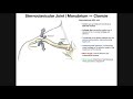
Sternoclavicular joint (Articulatio sternoclavicularis); Image: Yousun Koh
Sternoclavicular joint (Articulatio sternoclavicularis)
The sternoclavicular joint is a connection of three articular surfaces; the sternal end of the clavicle, the clavicular notch of the manubrium of sternum and the superior surface of the first costal cartilage.
The clavicular and sternal joint surfaces are convex and concave, respectively. These curvatures are found in the vertical plane. In the horizontal plane the articular surface of the clavicle is slightly concave while that of the sternum is convex, thus forming a sellar or saddle joint. The inferior aspect of both the clavicle and sternum, as well as the intervening joint space, rest on the first costal cartilage.
The radius of these respective curvatures differs, making the articular surfaces incongruent. The sternal end of the clavicle is also larger in size than the clavicular notch of the sternum, thus the medial end of the clavicle juts out superiorly above the upper border of the manubrium.
Joint congruency is enhanced somewhat by the presence of a fibrocartilaginous intra-articular disc. This disc lies between the clavicular and sternal surfaces, completely separating the joint into two compartments. The disc is held in place by several attachments, which strongly anchor it to the posterosuperior margin of clavicular articular surface, the costal cartilage of first rib, and to the joint capsule. The disc is the thinnest in its central part, which may perforate later in life.
Ligaments and joint capsule
The joint is surrounded by a fibrous joint capsule which is thickened on its anterior and posterior aspects but loose superiorly and inferiorly. The articular surfaces are lined with fibrocartilage.
Due to the lack of bony congruence, joint stability is provided by two sets of ligaments and the intra-articular disc. The ligaments are divided into;
Intrinsic ligaments; anterior and posterior sternoclavicular ligaments
Extrinsic ligaments; interclavicular and costoclavicular ligaments
The broad anterior sternoclavicular ligament runs from the anterosuperior surface of the sternal end of the clavicle to the anterosuperior surface of the manubrium and adjacent part of the first costal cartilage. This provides strong reinforcement to the anterior aspect of the joint. Similarly, the posterior sternoclavicular ligament covers the posterior aspect of the joint surfaces. Whilst also being broad, it is weaker than its anterior counterpart.
The interclavicular ligament emerges from the deep cervical fascia and connects the sternal ends of both clavicles, spanning the jugular notch. It strengthens the superior aspect of the joint capsule. The costoclavicular ligament unites the superior surface of the first rib and its costal cartilage with the inferomedial aspect of the clavicle. This strong thick ligamentous band consists of two sheaths (laminae) which differ in the direction of their fibers. The anterior lamina courses superolaterally, while the posterior fibers are directed superomedially, forming an inverted cone or cruciate shape. The costoclavicular ligament reinforces the inferior aspect of the joint, acting to limit clavicle elevation and anteroposterior movement.
Innervation
The sternoclavicular joint is innervated from two sources; superficially by the medial supraclavicular nerve (C3-C4; cervical plexus) and deeply by the nerve to subclavius (C5-C6; brachial plexus).
Blood supply
Blood supply to the sternoclavicular joint comes from branches of the suprascapular and internal thoracic arteries.
Movements
Due to the shape of its articular surfaces, the sternoclavicular joint is classified as a saddle joint. However, functionally, it has the features of a ball-and-socket joint, being a multiaxial joint with three degrees of freedom (mean active RoM values);
Elevation – depression (40°)
Protraction – retraction (35°)
Axial rotation (20-40°)
Sternoclavicular (SC) joint movements use the lateral end of the clavicle as a reference point. So during SC elevation the lateral end of the clavicle elevates, while during depression the lateral end of the clavicle lowers. In protraction the lateral end of the clavicle moves anteriorly while in retraction it moves posteriorly.
Видео Sternoclavicular Joint | Synovial Joint | Type | Attachments | Ligaments | clinical канала Knowing Anatomy
The function of the sternoclavicular joint is to coordinate the movements of the upper limb with the core of the body. Thus allowing the upper limb to perform its full range of motion. Specifically, the movements of the sternoclavicular joint are sorted into three degrees of freedom; elevation - depression, protraction - retraction, and axial rotation.
Articular surfaces
Sternoclavicular joint (Articulatio sternoclavicularis); Image: Yousun Koh
Sternoclavicular joint (Articulatio sternoclavicularis)
The sternoclavicular joint is a connection of three articular surfaces; the sternal end of the clavicle, the clavicular notch of the manubrium of sternum and the superior surface of the first costal cartilage.
The clavicular and sternal joint surfaces are convex and concave, respectively. These curvatures are found in the vertical plane. In the horizontal plane the articular surface of the clavicle is slightly concave while that of the sternum is convex, thus forming a sellar or saddle joint. The inferior aspect of both the clavicle and sternum, as well as the intervening joint space, rest on the first costal cartilage.
The radius of these respective curvatures differs, making the articular surfaces incongruent. The sternal end of the clavicle is also larger in size than the clavicular notch of the sternum, thus the medial end of the clavicle juts out superiorly above the upper border of the manubrium.
Joint congruency is enhanced somewhat by the presence of a fibrocartilaginous intra-articular disc. This disc lies between the clavicular and sternal surfaces, completely separating the joint into two compartments. The disc is held in place by several attachments, which strongly anchor it to the posterosuperior margin of clavicular articular surface, the costal cartilage of first rib, and to the joint capsule. The disc is the thinnest in its central part, which may perforate later in life.
Ligaments and joint capsule
The joint is surrounded by a fibrous joint capsule which is thickened on its anterior and posterior aspects but loose superiorly and inferiorly. The articular surfaces are lined with fibrocartilage.
Due to the lack of bony congruence, joint stability is provided by two sets of ligaments and the intra-articular disc. The ligaments are divided into;
Intrinsic ligaments; anterior and posterior sternoclavicular ligaments
Extrinsic ligaments; interclavicular and costoclavicular ligaments
The broad anterior sternoclavicular ligament runs from the anterosuperior surface of the sternal end of the clavicle to the anterosuperior surface of the manubrium and adjacent part of the first costal cartilage. This provides strong reinforcement to the anterior aspect of the joint. Similarly, the posterior sternoclavicular ligament covers the posterior aspect of the joint surfaces. Whilst also being broad, it is weaker than its anterior counterpart.
The interclavicular ligament emerges from the deep cervical fascia and connects the sternal ends of both clavicles, spanning the jugular notch. It strengthens the superior aspect of the joint capsule. The costoclavicular ligament unites the superior surface of the first rib and its costal cartilage with the inferomedial aspect of the clavicle. This strong thick ligamentous band consists of two sheaths (laminae) which differ in the direction of their fibers. The anterior lamina courses superolaterally, while the posterior fibers are directed superomedially, forming an inverted cone or cruciate shape. The costoclavicular ligament reinforces the inferior aspect of the joint, acting to limit clavicle elevation and anteroposterior movement.
Innervation
The sternoclavicular joint is innervated from two sources; superficially by the medial supraclavicular nerve (C3-C4; cervical plexus) and deeply by the nerve to subclavius (C5-C6; brachial plexus).
Blood supply
Blood supply to the sternoclavicular joint comes from branches of the suprascapular and internal thoracic arteries.
Movements
Due to the shape of its articular surfaces, the sternoclavicular joint is classified as a saddle joint. However, functionally, it has the features of a ball-and-socket joint, being a multiaxial joint with three degrees of freedom (mean active RoM values);
Elevation – depression (40°)
Protraction – retraction (35°)
Axial rotation (20-40°)
Sternoclavicular (SC) joint movements use the lateral end of the clavicle as a reference point. So during SC elevation the lateral end of the clavicle elevates, while during depression the lateral end of the clavicle lowers. In protraction the lateral end of the clavicle moves anteriorly while in retraction it moves posteriorly.
Видео Sternoclavicular Joint | Synovial Joint | Type | Attachments | Ligaments | clinical канала Knowing Anatomy
Показать
Комментарии отсутствуют
Информация о видео
Другие видео канала
 The Sternoclavicular (SC) Joint | Anatomy and Function
 Synovial Joints
 Clinical Anatomy - Knee mensicus and knee joint
 Muscles of Upper Arm | Two Compartments | Attachments | Origin | Insertion | Nerve Supply | Actions
 Elbow Joint: Bones, Muscles & Movement - Human Anatomy | Kenhub
 Hip bony bits, pelvis and ligaments
 Sternoclavicular Joint Dislocation Review - Everything You Need To Know - Dr. Nabil Ebraheim
 HUMERUS - GENERAL FEATURES & ATTACHMENTS
 The Acromioclavicular (AC) Joint | Anatomy and Function
 Clavicle Anatomy | General features, Osteology, Attachments, Development, clinical anatomy
 Shoulder joint Anatomy - Ligaments, Movements, Blood supply , Nerve supply and Clinical anatomy
 Radio Ulnar Joints | Anatomy | Upper limb
 LUNGS | ANATOMY | SIMPLIFIED
 Clinical Anatomy - Knee
 Scapulothoracic joint: Muscles and nerves
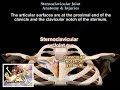 Anatomy Of The Sternoclavicular Joint - Everything You Need To Know - Dr. Nabil Ebraheim
 The 6 Types of Joints - Human Anatomy for Artists
 Shoulder Joint - 1 Feature and Ligament
 Synovial Joint Types
 Sternoclavicular Joint Dislocation Management | Orthopedic Classes