kidney and posterior abdominal wall - plastic model
Objectives:
After completion of this video session it is expected that you will be able to:
-- Describe the anterior and posterior relations of the kidney; relations of structures at the renal hilus.
-- Describe the shape and position of the suprarenal glands.
-- List the muscles of the posterior abdominal wall: psoas major, iliacus and quadratus lumborum. Summarize the attachments.
-- Classify the branches of abdominal aorta and list the distribution, branches and vertebral level of each class.
-- Describe the formation and course of the inferior vena cave; summarize its main tributaries.
-- Enumerate the sites of normal ureteric constrictions.
-- Describe the position of the kidneys and indicate the reason why the inferior pole of the right kidney is normally palpable.
Also check the related video: Kidney and posterior abdominal wall - dissection http://www.youtube.com/watch?v=E9JaJIhDIOI&feature=share
Presented and edited by Dr. Akram Jaffar (PhD). Filmed by Parwiz Akbari (medical student). Filmed at College of Medicine/ University of Sharjah, UAE. 2012.
This video and its channel are supported by "Human Anatomy Education" page on Facebook http://www.facebook.com/AnatomyEducation
Видео kidney and posterior abdominal wall - plastic model канала Human Anatomy Education
Показать
Комментарии отсутствуют
Информация о видео
Другие видео канала
 Posterior abdominal wall muscles
 Visceral Relations of the Kidney
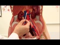 The Anatomy of the Ureter
 Kidney anatomy
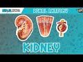 Renal | Kidney Anatomy Model
 Location and Relations of the Kidney - 3D Anatomy Tutorial
 Abdominal organs (plastic anatomy)
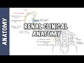 Kidneys - Clinical Anatomy (renal anatomy)
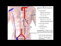 Blood Supply to the Anterior Abdominal Wall
 Muscles of the Thorax & Abdomen | Anatomy Model
 Lumbosacral plexus
 Renal system 3, Renal blood vessels
 Neurology - Spinal Cord Introduction
 Anatomy of the Liver
 Anatomy of the PERITONEUM || Dr. Yusuf ||
 Respiratory System 2, Upper airways
 Kidney - Sectional Anatomy
 Veins of the body - PART 2 - Anatomy Tutorial
 Gallstones Signs & Symptoms, Why They Occur | Cholecystitis, Choledocholithiasis, Cholangitis
 ANAT1010_25_Heart