Luciferase-Modified Magnetic Nanoparticles in Medical Imaging
By Philip Renkert, Emily Chen, and Megan Mazzatenta
Bioluminescent animals such as fireflies and dinoflagellates produce natural light in order to find mates and to protect their species from predators, but that same light has potential to greatly improve current medical practices. Medical imaging is crucial for tracking the progress of disease throughout the human body, but the synthesized chemicals currently used in the process inflict additional harm on the body, leaving scientists searching for a less invasive imaging method. By processing magnetic nanoparticles that are coated in luciferase, the main enzyme involved in bioluminescence, cells can be individually tracked in a natural, non-toxic manner.
In animals, agitation from the environment creates a flow of protons that bring the enzyme luciferase in contact with the compound luciferin. The luciferin is then oxidized in the presence of other compounds, such as adenosine triphosphate, and photons are released to generate light. Luciferase can be applied to track and image the growth of diseases, like cancer, noninvasively. Current medical imaging often requires the use of radiology, but because these techniques involve large amounts of radiation, there is often damage to the body. The light produced from the luciferase reaction is what gives the magnetic nanoparticles the ability to expose abnormal cell growth patterns, eliminating the need for other light sources that could harm the cells. A sensitive charge-coupled device camera can then be used to create a clear picture of the growth patterns by detecting the position of emitted light over time.
In processing luciferase for potential medical use in animals and humans, superparamagnetic iron oxide magnetic nanoparticles must first be synthesized by precipitating magnetite from a solution of ferric and ferrous ions. A polymer layer is added to prevent the particles from aggregating, and then the luciferase shell is added. The resultant processed MNP has a core with cubic spinel structure and is 40-119 nanometers in diameter. The superparamagnetic property of the nanoparticles can allow for cell manipulation by an external magnetic field. The cells targeted by certain nanoparticles can then be neutralized to lose function or separated from healthy ones. The nanoscale of the particles also allows for the tracking and manipulation of each cell individually, which can be done in a natural, safe way using the luciferase-modified MNPs.
In this video, we will explore the applications and possible drawbacks of the use of luciferase-modified magnetic nanoparticles in the medical imaging field.
References:
1. Cancer Gene Therapy and Cell Therapy. American Society of of Gene & Cell Therapy. http://www.asgct.org/general-public/educational-resources/gene-therapy-and-cell-therapy-for-diseases/cancer-gene-and-cell-therapy
2. Dikmen, Z. G. A New Diagnostic System in Cancer Research: Bioluminescent Imaging (BLI). TUBITAK, 35 (2005) 65-70. http://journals.tubitak.gov.tr/medical/issues/sag-05-35-2/sag-35-2-1-0404-18.pdf
3. Gupta, A. K.; Gupta, M. Synthesis and surface engineering of iron oxide nanoparticles for biomedical applications. Biomaterials 2005, 26(18), 3995–4021 DOI: 10.1016/j.biomaterials.2004.10.012.
4. Kocher, B.; Piwnica-Worms, D. Illuminating Cancer Systems With Genetically-Engineered Mouse Models and Coupled Luciferase Reporters In Vivo. Cancer discovery 2013, 3(6) 616-629 DOI: 10.1158/2159-8290.CD-12-0503.
5. Meroni, G.; Rajabi, M.; Santaniello, E. D-Luciferin, derivatives and analogues: synthesis and in vitro/in vivo luciferase-catalyzed bioluminescent activity. ARKIVOC 2009, 265-288.
6. Okabe, Y. Quantum dots for in vivo imaging: Disadvantages of Quantum Dots. University of California, Irvine. http://bme240.eng.uci.edu/students/07s/yokabe/disadvantages.htm
7. Roura, S.; Gálvez-Montón, C.; Bayes-Genis, A. Bioluminescence imaging: a shining future for cardiac regeneration. Journal of Cellular and Molecular Medicine 2013, 17(6) 693-703 DOI: 10.1111/jcmm.12018.
8. Sadikot, R.T.; Blackwell, T.S. Bioluminescence Imaging. Proceedings of the American Thoracic Society 2005, 2(6) 537-540 DOI: 9.1513/pats.200507-067DS.
9. Vasquez, E. S.; Feugang, J. M.; Willard, S. T.; Ryan, P. L.; Keisha, W. B. Journal of Nanobiotechnology 2016, 14 (20), DOI: 11.1186/s12951-016-0168-y.
10. Xu, T.; Close, D.; Handagama W.; Marr E.; Sayler G.; Ripp S. The Expanding Toolbox of In Vivo Bioluminescent Imaging. Frontiers in Oncology 2016, 6(150) DOI: 10.3389/fonc.2016.00150.
11. Zborowski, M.; Chalmers, J. J.; Lowrie, W. G. Magnetic Cell Manipulation and Sorting. Microsystems and Nanosystems Microtechnology for Cell Manipulation and Sorting 2016, 15–55 DOI: 10.1007/978-3-319-44139-9_2.
Sources: Images, Video Clips, and Audio
https://docs.google.com/document/d/1InA9tefQ5mZCnbMcSkbrKRliOfl3JUVrnT8mKfRGDGk/edit?usp=sharing
Видео Luciferase-Modified Magnetic Nanoparticles in Medical Imaging канала Intro to Materials Science--Guided Inquiry (UVa)
Показать
Комментарии отсутствуют
Информация о видео
10 мая 2017 г. 9:45:27
00:07:04
Другие видео канала
 Seeing the Smallest Thing in the Universe
 Bioluminescence: Don't Trust The Light
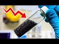 What's Graphene And Why It'll Soon Take Over The World
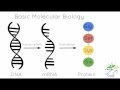 Luciferase Reporters in Circadian Research
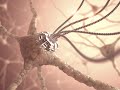 Nanotechnology: Hacking Humans, Its Potential, and Real Risks
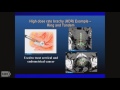 TRACO 2016: Radiation oncology and Small molecules
 Hydrogen - the Fuel of the Future?
 Magnetic Nanoparticles
 Biotechnology/Nanotechnology | Andrew Hessel | SingularityU Germany Summit 2017
 Luciferase Reporters for Mammals in Circadian Research
 Nanorobots in the Body: The Future of Medicine
 What Does An Atom REALLY Look Like?
 If You Get COVID 19: Optimize Immune System (Vitamin D, Monoclonal Antibodies, NAC, Quercetin etc.)
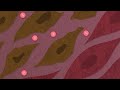 What are Magnetic Nanoparticles? | #Nanoscience featuring Diana Borca
 The technology behind the new COVID-19 mRNA vaccines
 Biotecnologie diagnostiche #2: metodi di sequenziamento del DNA (prof. Daniele Condorelli)
![1 A.M Study Session 📚 - [lofi hip hop/chill beats]](https://i.ytimg.com/vi/lTRiuFIWV54/default.jpg) 1 A.M Study Session 📚 - [lofi hip hop/chill beats]
1 A.M Study Session 📚 - [lofi hip hop/chill beats] COVID-19 RNA vaccines and the critical role of lipid nanoparticles
 Living Energy: Bioluminescent Bacteria in Solar Cells | Smiti Gandhi | TEDxClearBrookHighSchool
 Luciferase