Cockroach Dissection ( আরশোলার ব্যবচ্ছেদ ) | Biology Practical | Alamgir Kabir | NDC | 2018
In this article we will discuss about the dissection of cockroach. Also learn about:- 1. The Alimentary System 2. Dissection of Salivary Apparatus 3. Dissection of Nervous System 4,Digestive 5. Dissection of Reproductive System.
Killing:
The cockroach is usually killed with chloroform. It can be killed successfully by drowning in water.
ADVERTISEMENTS:
Dissection:
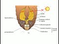
Hold the specimen (Fig. 6.1) with your left hand and clip the wings. Fix the specimen in a dorsal position on a dissecting tray with the help of pins passing through abdominal sterna and coxa of legs. Cut the lateral membrane (pleura) between the terga and sterna of the thorax and abdomen with a pair of fine scissors.
Posteriorly the two incisions should meet at the hindmost end of the abdomen. Proceed forward up to the anterior end of the thorax.
Give a transverse incision along the anterior border of the first thoracic segment and carefully remove the terga. The thoracic and the abdominal cavity are exposed. Put clear water in the tray. Remove fat bodies and tracheae to expose internal organs.
Mouth:
Ventral in position, located at the base of the buccal cavity.
Buccal cavity:
An ill-defined chamber, bounded anteriorly by epipharynx and labrum; posteriorly by hypo-pharynx and labium; laterally by two mandibles (Fig. 6.5).
Pharynx:
A short, vertically oriented tube opening into the oesophagus.
Oesophagus:
ADVERTISEMENTS:
A short tube running posteriorly in the thorax.
Crop:
There is no demarcation between the oesophagus and the crop. In fact, the crop is a large sac-like dilatation of the oesophagus. It extends into the abdomen.
Gizzard or proventriculus:
A round, thick walled muscular structure, posterior to the crop. It has two parts, the anterior contains six chitinous teeth in the inner wall and the posterior two circular hairy cushions.
Mesenteron or mid gut:
A narrow tube running from the gizzard to the hind gut. 7 to 8 hepatic caeca are present at the junction of the gizzard and mid gut.
Proctodaeum or hind gut:
A narrow tube, divisible into 3 zones – ileum, colon and rectum. The junction of the mid and hind gut is marked by 60 to 70 extremely fine, yellowish Malpighian tubules (The tubules are excretory in function.). The rectum opens through the anus.
Salivary glands:
Two in number. The glands and the receptacles lie on the dorsolateral aspects of the crop (Fig. 6.2). The ducts of the glands and receptacles run forward by the sides of the crop. The ducts from the two glands unite and those from the receptacles also unite to form two common ducts, which again unite and give rise to an efferent salivary duct opening on the ventral side of the hypo-pharynx.
Dissection of Salivary Apparatus:
Carefully remove all the tracheae and fat in the region where the salivary apparatus is lodged. Turn the crop as required and trace the ducts anteriorly running from the glands and the receptacles along the sides of the crop and then ventral to the oesophagus. Pin down the head of the cockroach with ventral surface upward.
Detach the glands and receptacles from the crop, separate the hypo-pharynx from the specimen by cutting it at the base and the salivary apparatus is free (Fig. 6.3). Carefully lift it with a spatula and place in a watch glass containing water. Mount it on a glass slide, if required.
Dissection of Nervous system...,
Circumoesophageal connectives:
Arising from the brain two short and broad nerves run around the oesophagus to meet the sub-oesophageal ganglia (Fig. 6.5).
Nervous System, Anterior Part, Lateral View
Sub-oesophageal ganglia:
It is formed by the fusion of two ganglia and located in the mid-ventral region of the head just ventral to the oesophagus. Nerves arising from sub-oesophageal ganglia end in labrum, mandible and both pairs of maxillae. A pair of connectives run backward from the ganglia and join the first thoracic ganglion, (prothoracic ganglion).
Ventral nerve cords:
These are two solid nerves and run along the mid-ventral line of the thorax and abdomen. The nerve cords are connected by nine ganglia, three thoracic and six abdominal.
The three thoracic ganglia and the last abdominal ganglion are large in size. Nerves emanating from the thoracic ganglia innervate the musculature and structures of the thoracic region. Those from the abdominal ganglia send nerves to the structures in the abdominal region.
Visceral Nervous System
i. Frontal ganglion:
Situated in front of brain, on the oesophagus, median in position. Connected to tritocerebral lobes of brain by a pair of frontal connectives.
ii. Hypo-cerebral ganglion:
Situated on the oesophagus below the brain. Recurrent nerve connects frontal and hypo-cerebral ganglion.
cockroach dissection ...
Видео Cockroach Dissection ( আরশোলার ব্যবচ্ছেদ ) | Biology Practical | Alamgir Kabir | NDC | 2018 канала MD. ALAMGIR KABIR
Killing:
The cockroach is usually killed with chloroform. It can be killed successfully by drowning in water.
ADVERTISEMENTS:
Dissection:
Hold the specimen (Fig. 6.1) with your left hand and clip the wings. Fix the specimen in a dorsal position on a dissecting tray with the help of pins passing through abdominal sterna and coxa of legs. Cut the lateral membrane (pleura) between the terga and sterna of the thorax and abdomen with a pair of fine scissors.
Posteriorly the two incisions should meet at the hindmost end of the abdomen. Proceed forward up to the anterior end of the thorax.
Give a transverse incision along the anterior border of the first thoracic segment and carefully remove the terga. The thoracic and the abdominal cavity are exposed. Put clear water in the tray. Remove fat bodies and tracheae to expose internal organs.
Mouth:
Ventral in position, located at the base of the buccal cavity.
Buccal cavity:
An ill-defined chamber, bounded anteriorly by epipharynx and labrum; posteriorly by hypo-pharynx and labium; laterally by two mandibles (Fig. 6.5).
Pharynx:
A short, vertically oriented tube opening into the oesophagus.
Oesophagus:
ADVERTISEMENTS:
A short tube running posteriorly in the thorax.
Crop:
There is no demarcation between the oesophagus and the crop. In fact, the crop is a large sac-like dilatation of the oesophagus. It extends into the abdomen.
Gizzard or proventriculus:
A round, thick walled muscular structure, posterior to the crop. It has two parts, the anterior contains six chitinous teeth in the inner wall and the posterior two circular hairy cushions.
Mesenteron or mid gut:
A narrow tube running from the gizzard to the hind gut. 7 to 8 hepatic caeca are present at the junction of the gizzard and mid gut.
Proctodaeum or hind gut:
A narrow tube, divisible into 3 zones – ileum, colon and rectum. The junction of the mid and hind gut is marked by 60 to 70 extremely fine, yellowish Malpighian tubules (The tubules are excretory in function.). The rectum opens through the anus.
Salivary glands:
Two in number. The glands and the receptacles lie on the dorsolateral aspects of the crop (Fig. 6.2). The ducts of the glands and receptacles run forward by the sides of the crop. The ducts from the two glands unite and those from the receptacles also unite to form two common ducts, which again unite and give rise to an efferent salivary duct opening on the ventral side of the hypo-pharynx.
Dissection of Salivary Apparatus:
Carefully remove all the tracheae and fat in the region where the salivary apparatus is lodged. Turn the crop as required and trace the ducts anteriorly running from the glands and the receptacles along the sides of the crop and then ventral to the oesophagus. Pin down the head of the cockroach with ventral surface upward.
Detach the glands and receptacles from the crop, separate the hypo-pharynx from the specimen by cutting it at the base and the salivary apparatus is free (Fig. 6.3). Carefully lift it with a spatula and place in a watch glass containing water. Mount it on a glass slide, if required.
Dissection of Nervous system...,
Circumoesophageal connectives:
Arising from the brain two short and broad nerves run around the oesophagus to meet the sub-oesophageal ganglia (Fig. 6.5).
Nervous System, Anterior Part, Lateral View
Sub-oesophageal ganglia:
It is formed by the fusion of two ganglia and located in the mid-ventral region of the head just ventral to the oesophagus. Nerves arising from sub-oesophageal ganglia end in labrum, mandible and both pairs of maxillae. A pair of connectives run backward from the ganglia and join the first thoracic ganglion, (prothoracic ganglion).
Ventral nerve cords:
These are two solid nerves and run along the mid-ventral line of the thorax and abdomen. The nerve cords are connected by nine ganglia, three thoracic and six abdominal.
The three thoracic ganglia and the last abdominal ganglion are large in size. Nerves emanating from the thoracic ganglia innervate the musculature and structures of the thoracic region. Those from the abdominal ganglia send nerves to the structures in the abdominal region.
Visceral Nervous System
i. Frontal ganglion:
Situated in front of brain, on the oesophagus, median in position. Connected to tritocerebral lobes of brain by a pair of frontal connectives.
ii. Hypo-cerebral ganglion:
Situated on the oesophagus below the brain. Recurrent nerve connects frontal and hypo-cerebral ganglion.
cockroach dissection ...
Видео Cockroach Dissection ( আরশোলার ব্যবচ্ছেদ ) | Biology Practical | Alamgir Kabir | NDC | 2018 канала MD. ALAMGIR KABIR
Показать
Комментарии отсутствуют
Информация о видео
Другие видео канала
 Female reproductive system and development of Cockroach for NEET AIIMS
 Cockroach Mouthparts Dessection ( আরশোলার মুখোপাঙ্গ ব্যবচ্ছেদ ) | Biology Practical
 Feeding (and Cleaning Up After) Our Fry | at the Leavenworth National Fish Hatchery
 Cockroach dissection - Digestive system
 5 Fish That have Worms in Their Meat
 Cockroach - Made Easy for You | NEET Biology | NEET 2022 | Ritu Rattewal
 Salmon Anatomy Part 2: Internal Anatomy
 Tips and Tricks to Remember Floral Formula - 1 | NEET 2020 | Unacademy NEET | Biology | Pradeep Sir
 Dissection of Ruhi fish(Taaki fish)|Bio-Practical|Class11-12
 The structure of bees' legs
 Dissection of Cockroach(americana)|Bio-practical|Class 11-12
 Observation of Mitosis in Onion Root tip Experiment | Practical, Procedure
 Dissection of cockroach performed by students | class 11th biology | Must Watch 🧐
 Laboratory Equipment/ Instrument Names, Meaning & Images | Laboratory Equipment Vocabulary
 01. হাইড্রার পরিচিতি । HSC Zoology Chapter 2 | Introduction to Hydra | Fahad Sir
 Cockroach: reproductive system - female || XI BIOLOGY || CBSE || NEET || AIIMS
 Salivary gland of Cockroach / Periplaneta americana
 Blood Cell Count: Using Haemocytometer
 Mounting prawn appendages for BSc Zoology students
 Digestive System of Pila ( Pila globosa) - শামুকের পরিপাকতন্ত্র পর্যবেক্ষণ