Ultimate guide to MR anatomy of the Posterolateral Corner in the Knee
How to assess the posterolatearal corner on MRI. In this video, I show you the anatomy of all posterolateral corner structures (PCL) that you need to know and how to simplify the assessment of knee MRI for posterolateral corner injuries. It does not have to be complicated.
The three important structure you need to assess are the:
1. lateral collateral ligament (LCL)
2. biceps femoris tendon
3. popliteus tendon
Smaller structures you can asses are:
4. poplitefibular ligament
5. meniscopopliteal fascicles
6. fabellofibular ligament
7. arcuate ligament
If you want to go deeper, check out this two selected scientific articles:
https://pubs.rsna.org/doi/10.1148/rg.2016160027
https://www.ncbi.nlm.nih.gov/pubmed/?term=filli+posterolateral+corner
#kneeMRI #PLC #MRI
Please subscribe to my channel and also check out my patreon page:
SUPPORT MY CHANNEL HERE:
https://www.patreon.com/agten
Patreon is a online system, where you can support me on a more personal level with a tiny donation in exchange for small benefits, as listed on my page. It is a great way to engage with me and learn together. Every month I post patreon-only videos over on my patreon page.
Thanks for watching! You need an MRI and want it analyzed by me personally? Go to www.aristra.com (Germany and Switzerland), also available on www.aristra.de and aristra.ch #ARISTRA
Sie brauchen ein MRT und wollen den Befund durch mich? Melden Sie sich zur MRT an auf www.aristra.de , dann kann ich Ihnen helfen :)
Видео Ultimate guide to MR anatomy of the Posterolateral Corner in the Knee канала Dr Christoph Agten
Показать
Комментарии отсутствуют
Информация о видео
Другие видео канала
 Get A First Look At Virtual MSK Fellowship Case Discussions In This Hour-long Video!
 MSK MRI Live Reporting Session (German/English)
 MSK MRI Rapid Fire Session - Ep.01
 Q&A with Dr Agten August 9, 2023
 Speed vs. Accuracy: Knee MRI Analysis Showdown!
![Radiologist at work - 10 MSK MRI in under 80min - LIVE reporting session [GERMAN]](https://i.ytimg.com/vi/g_5U2xou4GM/default.jpg) Radiologist at work - 10 MSK MRI in under 80min - LIVE reporting session [GERMAN]
Radiologist at work - 10 MSK MRI in under 80min - LIVE reporting session [GERMAN] ESSR 2023 Bilbao Highlight 2 - Anterior Capsular Ligament
 ESSR 2023 Bilbao Highlight 1 - Lateral Ankle Ligament Variants
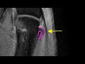 Thumb MRI: Stener-like lesion of the RADIAL collateral ligament
 The Bridging Sign In Rotator Cuff Tears on Shoulder MRI
 MRI Triceps Tendon Injury and Anatomy
 How much time do radiologists save by using A.I. tools?
 ChatGPT wrote text: How to Increase Confidence in Reporting MSK MRI
 MRI of DRUJ Instability / Distal radioulnar joint instability measurement
 How to use ChatGPT in Radiology
 Testimonial Virtual MSK Fellowship Program
 MRI of glenoid labral flap tear - a pathognomonic lesion (rare)
 plantar vein thrombosis
 MRI in Axial Spondylarthritis: Thoracic Spine Inflammation (MSK MRI Quick Tip 7)
 Philips PSM6300 SpeechOne Wireless Headset Unboxing and Setup
 Best Microphone for Radiology Speech Recognition for Home office and Teleradiology