Pharynx Anatomy (Parts, Layers, Muscles)
Content:
Introduction 0:00
Parts of the Pharynx: 00:36
Nasopharynx: 01:53
Oropharynx: 07:19
Laryngopharynx: 07:44
Layers of Pharyngeal Wall: 08:36
Muscles of the Pharynx 10:17
-------------------------------
💎Channel membership: https://www.youtube.com/channel/UCEr7pkSXVsHcBLLBcJAGV-Q/join
📷 Follow my IG: https://www.instagram.com/taimtalksmed/
💝 Donation link: https://www.buymeacoffee.com/taimtalksmed
-------------------------------
Parts of the Pharynx:
- Nasopharynx, Oropharynx, Laryngopharynx
Layers of the Pharyngeal Wall
Muscles of the Pharynx
Pharynx:
- 12 to 15 cm long
- Nasopharynx (Pars Nasalis)
- Oropharynx (Pars Oralis)
- Laryngopharynx (Pars Laryngis)
Nasopharynx:
- Level of C1-C2
- Vault of Pharynx (Fornix Pharyngis)
- Attachment points of the pharynx: Pharyngeal Tubercle of the occipital bone (tuberculum pharyngeum), Petrooccipital Fissure (petrooccipital synchondrosis), Inferior Surface of Petrous Part (Temporal Bone), Medial Lamina of Pterygoid Process
- Choana (Internal Nose)
- Auditory Tube
- Pharyngeal opening of the auditory tube (Ostium Pharyngeum Tubae Auditiva)
- Cushion of the Auditory Canal (Torus Tubarius)
- Pharyngeal Recess (Recessus Pharyngeus)
- Pharyngeal Tonsils / Adenoids (Tonsilla pharyngealis)
- Tubal Tonsils (Tonsilla Tubaria)
Overview of Auditory Tube:
- Ear Structures: Outer Ear, Middle Ear (ossicles and tympanic membrane), Inner ear.
- Eustachian Tube connects the nasopharynx with the middle ear
- Function of auditory tube:
○ Equalizing the pressure
○ Draining the middle ear
Oropharynx:
- Level of C4-C4
- Bordered by Soft Palate and Epiglottis
- Connects with the Oral cavity through the Oropharyngeal Isthmus (Isthmus faucium)
Laryngopharynx:
- Level of C5-C6
- Continues into Larynx through Laryngeal Inlet (auditus laryngis)
- Piriform Fossa (Recessus Piriformis)
Layers of the Pharyngeal Wall:
- Tunica Mucosa
○ Lined by Respiratory Epithelium (Pseudostratified epithelium with cilia and goblet cells)
○ Lined by Stratified Squamous non keratinized epithelium
- Tela Submucosa
○ Contains Connective tissue with blood vessels and lymph vessels, and glands
- Tunica Muscularis
○ Has 2 muscle layers for peristalsis
○ Internal Circular muscle layer (Stratum Circulare)
○ Outer Longitudinal Muscle layer (Stratum longitudinale)
- Tunica Adventitia
○ Covers Pharynx from outside
External Pharyngeal Muscles:
- Pharyngeal Constrictors (Musculi Constrictores Pharyngis)
○ Superior Pharyngeal Constrictor
○ Medial Pharyngeal Constrictor
○ Inferior Pharyngeal Constrictor
- Pharyngeal Elevators (Musculi Levatores Pharyngis)
○ Stylopharyngeus Muscle (Musculus Stylopharyngeum)
○ Palatopharyngeus Muscle (Musculus Palatopharyngeum)
○ Salpingopharyngeus Muscle (Musculus Salpingopharyngeus)
Sources used in this video:
- Memorix Anatomy 2nd Edition by Hudák Radovan (Author), Kachlík David (Author), Volný Ondřej (Author)
- Biorender
- University notes and lectures
Видео Pharynx Anatomy (Parts, Layers, Muscles) канала Taim Talks Med
Показать
Комментарии отсутствуют
Информация о видео
Другие видео канала
 Introducing Taim Talks Cardio: New Animation!
 Neuron (Membrane Potentials, Action Potential, All-or-None Law) | Physiology
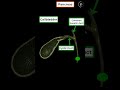 Gall bladder and Pancreas in 1 min
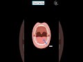 Oral Cavity in 1 min
 CN 6: Abducens Nerve (Scheme, Pathway, Clinical Relevance) | Neuroanatomy
 Announcing 1-minute videos
 Topography of the Hip (Foramina, Canals, Spaces, +Femoral Canal)
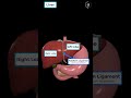 Liver in 1 min
 Fascia of the Thorax (Endothoracic, Thoracic, Clavipectoral, Pectoral Fascia)
 Minor & Major Salivary Glands (Parotid, Submandibular, Sublingual) Anatomy
 Salivary Glands in 1 min
 Stomach in 1 min
 Topography of the Thigh and Leg (Femoral Triangle, Adductor Canal, Popliteal Fossa)
 Cellular Immunity (Steps, Immunity to Viruses and Interferons) | Immunology
 CN 11: Accessory Nerve (Scheme, Nuclei, Pathway, Branches) | Neuroanatomy
 Parasympathetic Nervous System (Overview, Scheme) | Neuroanatomy
 Fascia of the Abdomen (Superficial, Investing Abdominal, Endoabdominal)
 Abdominal Aorta (Branches + Mnemonics)
 Portal Venous System & Porto-Caval Anastomosis (Tributaries and Clinical Significance) - Anatomy
 Why I Changed My Youtube Name | Meditay
 CN 4: Trochlear Nerve (Scheme, Pathway, Clinical Relevance) | Anatomy/Neurology