Anatomy Of The Patellar Tendon - Everything You Need To Know - Dr. Nabil Ebraheim
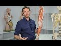
Dr. Ebraheim’s educational animated video describes the anatomy of the Patellar tendon.
The patellar tendon attaches the patella to the top of the tibia. The quadriceps muscle is attached superiorly to the patella. A small part of the quadriceps tendon then continues over the front of the patella to become the patellar tendon.
Diagram showing the forces and constraints applied to the patella during function.
Arterial supply of the patellar tendon:
•Descending genicular artery
•Superior medial genicular artery
•Superior lateral genicular artery
•Circumflex fibular artery
•Anterior tibial artery
•Inferior medial genicular artery.
The patellar tendon works with the quadriceps to straighten the leg.
Several bursae are seen around the patella
•Suprapatellar
•Prepatellar
•Infrapatellar
These bursae allow the knee cap to slide freely underneath the skin while bending and straightening the knee. The bursa may become inflamed due to trauma or infection, however, bursitis of the knee most commonly occurs over the kneecap.
The femoral condyles are covered with hyaline cartilage and form the femoral trochlea. The patella articulates with the femoral trochlea. The patella lies just above the femoral trochlea when the knee is in full extension. The patella is classified as a sesamoid bone of the quadriceps tendon with a proximal base and a distal apex (triangular). The lateral facet is larger than the medial facet. The articular surface of the patella is covered with hyaline cartilage and has two articular facets for the femur. The apex which is the distal part is nonarticular. This area does give attachment to the patellar tendon.
Patellar tendonitis may develop due to repeated stress being placed on the patellar tendon. This condition occurs in athletes from overuse. A weakened patellar tendon is more likely to tear and it may become torn when it attached to the kneecap. Patellar tendon tears can be either partial or complete. When the patellar tendon is ruptured, the quadriceps will pull the patella upward. Imaging tests such as an x-ray or MRI may be ordered to confirm the presence of a patellar tendon rupture. Complete tears can often be identified by x-ray alone. MRI showing rupture of the patellar tendon.
One way to measure the patellar height is by measuring the Blumensaat’s line. The knee needs to be flexed at least 30 degrees then a line can be drawn through the roof of the intercondylar notch and usually touches the tip of the patella.
The patella moves upward with the patellar tendon rupture (patella alta). Rupture of the quadriceps tendon causes patella Baja (infra). Lateral dislocation of the patella is also seen in “sunrise” view. May also cause direct impact injury to the medial patella and lateral femoral condyle. May also cause direct impact and injury to the medial patella and lateral femoral condyle.
Become a friend on facebook:
http://www.facebook.com/drebraheim
Follow me on twitter:
https://twitter.com/#!/DrEbraheim_UTMC
Видео Anatomy Of The Patellar Tendon - Everything You Need To Know - Dr. Nabil Ebraheim канала nabil ebraheim
Показать
Комментарии отсутствуют
Информация о видео
Другие видео канала
 The Patella (anatomy)
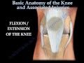 Knee injury ,Injuries - Everything You Need To Know - Dr. Nabil Ebraheim
 Patella Tendon Repair
 Why You Have a Kneecap and How it Unleashes Your Quads
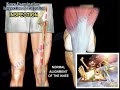 Knee Examination Inspection & Palpation - Everything You Need To Know - Dr. Nabil Ebraheim
 LIGAMENTS OF THE KNEE
 Tests For Examination Of The Knee - Everything You Need To Know - Dr. Nabil Ebraheim
 Basic Anatomy Of The Patella - Everything You Need To Know - Dr. Nabil Ebraheim
 Knee Pain Types Explained | Royersford, PA | Limerick, PA
 Quadriceps Tendon Rupture - Everything You Need To Know - Dr. Nabil Ebraheim
 Clinical Anatomy - Knee
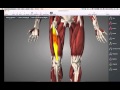 Muscles that move knee
 Knee pain above the patella | Physiotherapy | Quad tendinopathy | PhysioEvangelist
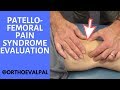 Patellofemoral Pain Syndrome Evaluation and Treatment
 Patellar Fractures - Everything You Need To Know - Dr. Nabil Ebraheim
 Knee Series (2 of 5): 9 Patellar Tendonitis Treatments/ Assessments
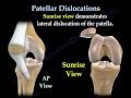 Patellar Dislocations - Everything You Need To Know - Dr. Nabil Ebraheim
 Rehab for patella tendon rupture and repair | Feat. Tim Keeley | No.38 | Physio REHAB
 How to Fix Patellar Tendonitis (No More KNEE PAIN!)
 Quadriceps Tendon Rupture - Everything You Need To Know - Nabil Ebraheim