- Популярные видео
- Авто
- Видео-блоги
- ДТП, аварии
- Для маленьких
- Еда, напитки
- Животные
- Закон и право
- Знаменитости
- Игры
- Искусство
- Комедии
- Красота, мода
- Кулинария, рецепты
- Люди
- Мото
- Музыка
- Мультфильмы
- Наука, технологии
- Новости
- Образование
- Политика
- Праздники
- Приколы
- Природа
- Происшествия
- Путешествия
- Развлечения
- Ржач
- Семья
- Сериалы
- Спорт
- Стиль жизни
- ТВ передачи
- Танцы
- Технологии
- Товары
- Ужасы
- Фильмы
- Шоу-бизнес
- Юмор
Amoebiasis – Transmission, Pathogenesis, Diagnosis & Treatment
Amoebiasis – Transmission, Pathogenesis, Diagnosis & Treatment
Amoebiasis – caused by the parasite Entamoeba histolytica – is a major cause of diarrheal disease and liver abscess worldwide, and a high-yield topic on every medical exam. In this video, THE NEXUS MEDICS takes you from micro to macro, covering the parasite biology, life cycle, transmission, pathogenesis (including the famous "flask-shaped" ulcer), intestinal and extraintestinal manifestations, diagnosis, treatment, and public health.
Causative Agent: Entamoeba histolytica – a protozoan parasite belonging to the phylum Amoebozoa. Distinguished from non-pathogenic Entamoeba dispar (morphologically identical but genetically distinct – does NOT cause disease) and Entamoeba moshkovskii. Only E. histolytica causes invasive disease. Other commensal amoebae: Entamoeba coli, Endolimax nana, Iodamoeba bütschlii (all non-pathogenic, different nuclear morphology).
Morphology (Two Forms):
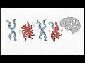
Cyst (Infectious Form): Spherical, 10-20μm, mature cyst contains 4 nuclei (chromatoidal bodies – cigar-shaped aggregates of ribosomes in young cysts). Resistant to gastric acid, chlorine, drying (survives days to weeks in environment). Transmitted via fecal-oral route.
Trophozoite (Invasive Form): Irregular, 20-40μm, single nucleus with central karyosome, ingested RBCs (erythrophagocytosis – diagnostic hallmark of pathogenic E. histolytica). Motile via pseudopodia. Replicates in colon. Does NOT survive outside host (dies rapidly).
Life Cycle: Human ingests mature cysts (contaminated food/water, fecally contaminated hands, vegetables fertilized with human feces) → cysts excyst in small intestine → each cyst releases 8 trophozoites → trophozoites migrate to large intestine (cecum, ascending colon, sigmoid colon – sites of predilection) → trophozoites multiply by binary fission → trophozoites invade colonic mucosa (if conditions favorable) OR encyst (form new cysts) in intestinal lumen → cysts excreted in feces (host sheds millions/day) → transmission to new host. No animal reservoir (humans are definitive host). No intermediate host.
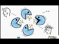
Pathogenesis (How It Causes Disease): Trophozoites adhere to colonic mucin and epithelial cells via Gal/GalNAc lectin (carbohydrate-binding protein) – virulence factor. Secretes amoebapores (pore-forming proteins that kill host cells) and cysteine proteases (degrade extracellular matrix – collagen, fibronectin, laminin, degrade IgA and complement). Host inflammatory response (neutrophils recruited but killed by trophozoites, releasing lysosomal enzymes that cause more tissue damage). Typical lesion: "flask-shaped" ulcer – narrow neck (mucosal breach) with wide base (submucosal undermining). Overlying mucosa may appear normal. Severe cases: diffuse colitis, toxic megacolon, amoeboma (granulomatous mass, can mimic carcinoma).
#Amoebiasis #EntamoebaHistolytica #NexusMedics #THEMEDICS #MicroToMacro #FlaskShapedUlcer #AmoebicDysentery #AmoebicLiverAbscess #AnchovyPaste #Metronidazole #Tinidazole #Paromomycin #LuminalAgent #CystVsTrophozoite #Erythrophagocytosis #GalGalNAcLectin #Amoebapore #CysteineProtease #FulminantColitis #ToxicMegacolon #Amoeboma #RightLobeAbscess #HepatobronchialFistula #PericardialAmoebiasis #NoFeverInColitis #TravelersDiarrhea #EndemicAreas #MSM #FecalOral #ContaminatedWater #StoolPCR #AntigenDetection #SerologyAmoebiasis #ColonoscopyFlaskUlcer #UltrasoundHypoechoic #CTDoubleTargetSign #AspirationNotRoutine #DiloxanideFuroate #Iodoquinol #RelapsePrevention #USMLEparasitology #COMLEXinfectiousdisease #NCLEXglobalhealth #PLABtropicalmedicine #MBBSprotozoa #NeglectedTropicalDisease #HealthAcademics #PublicHealth #NexusMethod #AmoebiasisTreatment
Видео Amoebiasis – Transmission, Pathogenesis, Diagnosis & Treatment канала The Nexus Medics
Amoebiasis – caused by the parasite Entamoeba histolytica – is a major cause of diarrheal disease and liver abscess worldwide, and a high-yield topic on every medical exam. In this video, THE NEXUS MEDICS takes you from micro to macro, covering the parasite biology, life cycle, transmission, pathogenesis (including the famous "flask-shaped" ulcer), intestinal and extraintestinal manifestations, diagnosis, treatment, and public health.
Causative Agent: Entamoeba histolytica – a protozoan parasite belonging to the phylum Amoebozoa. Distinguished from non-pathogenic Entamoeba dispar (morphologically identical but genetically distinct – does NOT cause disease) and Entamoeba moshkovskii. Only E. histolytica causes invasive disease. Other commensal amoebae: Entamoeba coli, Endolimax nana, Iodamoeba bütschlii (all non-pathogenic, different nuclear morphology).
Morphology (Two Forms):
Cyst (Infectious Form): Spherical, 10-20μm, mature cyst contains 4 nuclei (chromatoidal bodies – cigar-shaped aggregates of ribosomes in young cysts). Resistant to gastric acid, chlorine, drying (survives days to weeks in environment). Transmitted via fecal-oral route.
Trophozoite (Invasive Form): Irregular, 20-40μm, single nucleus with central karyosome, ingested RBCs (erythrophagocytosis – diagnostic hallmark of pathogenic E. histolytica). Motile via pseudopodia. Replicates in colon. Does NOT survive outside host (dies rapidly).
Life Cycle: Human ingests mature cysts (contaminated food/water, fecally contaminated hands, vegetables fertilized with human feces) → cysts excyst in small intestine → each cyst releases 8 trophozoites → trophozoites migrate to large intestine (cecum, ascending colon, sigmoid colon – sites of predilection) → trophozoites multiply by binary fission → trophozoites invade colonic mucosa (if conditions favorable) OR encyst (form new cysts) in intestinal lumen → cysts excreted in feces (host sheds millions/day) → transmission to new host. No animal reservoir (humans are definitive host). No intermediate host.
Pathogenesis (How It Causes Disease): Trophozoites adhere to colonic mucin and epithelial cells via Gal/GalNAc lectin (carbohydrate-binding protein) – virulence factor. Secretes amoebapores (pore-forming proteins that kill host cells) and cysteine proteases (degrade extracellular matrix – collagen, fibronectin, laminin, degrade IgA and complement). Host inflammatory response (neutrophils recruited but killed by trophozoites, releasing lysosomal enzymes that cause more tissue damage). Typical lesion: "flask-shaped" ulcer – narrow neck (mucosal breach) with wide base (submucosal undermining). Overlying mucosa may appear normal. Severe cases: diffuse colitis, toxic megacolon, amoeboma (granulomatous mass, can mimic carcinoma).
#Amoebiasis #EntamoebaHistolytica #NexusMedics #THEMEDICS #MicroToMacro #FlaskShapedUlcer #AmoebicDysentery #AmoebicLiverAbscess #AnchovyPaste #Metronidazole #Tinidazole #Paromomycin #LuminalAgent #CystVsTrophozoite #Erythrophagocytosis #GalGalNAcLectin #Amoebapore #CysteineProtease #FulminantColitis #ToxicMegacolon #Amoeboma #RightLobeAbscess #HepatobronchialFistula #PericardialAmoebiasis #NoFeverInColitis #TravelersDiarrhea #EndemicAreas #MSM #FecalOral #ContaminatedWater #StoolPCR #AntigenDetection #SerologyAmoebiasis #ColonoscopyFlaskUlcer #UltrasoundHypoechoic #CTDoubleTargetSign #AspirationNotRoutine #DiloxanideFuroate #Iodoquinol #RelapsePrevention #USMLEparasitology #COMLEXinfectiousdisease #NCLEXglobalhealth #PLABtropicalmedicine #MBBSprotozoa #NeglectedTropicalDisease #HealthAcademics #PublicHealth #NexusMethod #AmoebiasisTreatment
Видео Amoebiasis – Transmission, Pathogenesis, Diagnosis & Treatment канала The Nexus Medics
amoebiasis Entamoeba histolytica Entamoeba dispar differentiation amoebiasis life cycle cyst infectious form 4 nuclei trophozoite invasive form erythrophagocytosis fecal-oral transmission amoebiasis contaminated water food amoeba Gal/GalNAc lectin virulence amoebapores pore-forming proteins cysteine proteases ECM degradation flask-shaped ulcer pathology amoebic colitis bloody diarrhea mucus tenesmus
Комментарии отсутствуют
Информация о видео
17 мая 2026 г. 23:50:14
00:08:35
Другие видео канала