Melanocytes vs Keratinocytes Made Easy: 5-Minute Pathology Pearls
A complete organized library of all my videos, digital slides, pics, & sample pathology reports is available here: https://kikoxp.com/posts/5084 (dermpath) & https://kikoxp.com/posts/5083 (bone/soft tissue sarcoma pathology).
Melanocytes and keratinocytes both can develop artifactual clear vacuoles around them. To dermpath beginners, this can make them difficult to tell apart. Once you know the trick, you can usually distinguish vacuolated melanocytes from vacuolated keratinocytes easily on H&E. And yes, I know this one is technically longer than 5 minutes. ;-)
Thanks to my former medical student, Sophia Ly, for typing up the transcript of this video so it could have accurate closed caption/subtitles! You can access a text version of her transcript of my video here: https://kikoxp.com/posts/5389
Other videos:
- Normal Skin Histology: https://youtu.be/yQQ2Dmz42Vs
- Melanoma Basics: https://youtu.be/8N0IZZpF8ts
This video is geared towards medical students, pathology or dermatology residents, or practicing pathologists or dermatologists. Of course, this video is for educational purposes only and is not formal medical advice or consultation.
Presented by Jerad M. Gardner, MD. Please subscribe to my channel to be notified of new pathology teaching videos.
Follow me on:
Snapchat: JMGardnerMD
Twitter: @JMGardnerMD
Instagram: @JMGardnerMD
Facebook: https://www.facebook.com/JMGardnerMD/
Видео Melanocytes vs Keratinocytes Made Easy: 5-Minute Pathology Pearls канала Jerad Gardner, MD
Показать
Комментарии отсутствуют
Информация о видео
Другие видео канала
 Melanocytic Dermpath Basics: Benign Nevus
 Keratin Pearl (SCC) vs Horn Pseudocyst (SK) (aka "pseudohorn cyst")
 Lifecycle of the Keratinocyte
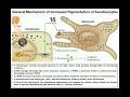 How do Melanocytes Make Melanin?: Melanogenesis Mechanism
 How We Get Our Skin Color | HHMI BioInteractive Video
 Histopathology Skin --Malignant melanom
 Immunohistochemistry in Normal Skin: Cytokeratins
 Atypical fibroxanthoma (AFX) vs mimics (spindle cell melanoma, squamous cell carcinoma, etc)
 Melanocytic Dermpath Basics: Melanoma
 Seborrheic Keratosis: 5-Minute Pathology Pearls
 Disseminated intravascular coagulation - causes, symptoms, diagnosis, treatment, pathology
 Ganglion Cyst...with an AMAZING Bonus! Beautiful Polarizable Maltese Cross Paraffin Crystals
 Syringocystadenoma Papilliferum (SCAP) 101...Explained by a Dermatopathologist
 Skin Adnexal Tumors: Dermatopathology Unknown Cases
 Squamous Cell Carcinoma & Actinic Keratosis 101...Dermpath Basics & Beyond
 Mycosis Fungoides (Cutaneous T-Cell Lymphoma): 5-Minute Pathology Pearls
 Dysplastic Nevus: 5-Minute Pathology Pearls
 Skin Adnexal Tumors 101: A Basic Approach for General Pathologists
 Inflammatory Dermpath 101 (A Beginner's Guide to Diagnosing Skin Rashes for Non-Dermatopathologists)
 IMT: Inflammatory Myofibroblastic Tumor...Explained by a Soft Tissue Pathologist