THE INFLAMMATORY RESPONSE
The inflammatory response is initiated within hours of infection or wounding and is characterized by edema, or swelling, heat, redness, and pain at the site of an infection or injury. These characteristics reflect four changes in local blood vessels.
1. The heat and redness during inflammation is the result of an increase in vascular diameter. The increase in vascular diameter also results in slower blood flow.
2. There is an increase in vascular permeability. During inflammation, endothelial cells have gaps between them - fluid from the blood exits and accumulates in local tissues, resulting in edema and pain. The fluid contains plasma proteins such as complement proteins and mannose binding lectin, which aid in defending against pathogens.
3. Endothelial cells, which line the walls of blood vessels, are “activated” during inflammation. That is, endothelial cells express cell-adhesion molecules that promote the binding of circulating leukocytes, otherwise known as white blood cells.
4. There is clotting in the microvessels at the site of infection, which prevents pathogens from spreading via the blood.
The purpose of the inflammatory response is threefold:
1. Allows the body to defend itself from invading microorganisms. The increase in vascular diameter, along with the activated endothelial cells, results in leukocytes being able to attach to the endothelium, and then migrate into the tissues where they can attack pathogens. This process of leukocytes leaving the bloodstream and entering tissues is called extravasation.
2. Induces local blood clotting, and this creates a physical barrier preventing the infection from spreading into the bloodstream.
3. Promotes the repair of injured tissue.
The state of inflammation is set up when tissues are physically damaged, or when pathogens are recognized by macrophages and later by other white blood cells. These circumstances induces the release of a variety of inflammatory mediators which cause the inflammatory response. Macrophages and neutrophils secrete prostaglandins, leukotrienes, and platelet-activating factor (PAF), which are lipid mediators of inflammation.
Then, macrophages secrete cytokines, which are substances released by cells of the immune system that affect other cells. One kind of cytokine are chemokines, which act as chemoattractants. Chemokines cause directed chemotaxis. Chemokines direct phagocytes to move towards the source of the chemokines, which are the sites where they are needed.
Two cytokines important to the inflammatory response are C5a and Tumor Necrosis Factor-α , or TNF-α. C5a stimulates respiratory burst, which is the rapid release of reactive oxygen species. It attracts neutrophils and monocytes. It also increases vascular permeability, increases expression of adhesion molecules on the endothelium, and causes local mast cells to release granules containing the inflammatory molecule histamine, and TNF-α. TNF-α is a potent activator of endothelial cells.
Activation of endothelial cells is central to the inflammatory response. Cytokines produced by macrophages, especially TNF-α, cause endothelial cells to externalize granules called Weibel-Palade bodies containing P-selectin within minutes of pathogen detection by macrophages. P-selectin appears on the surfaces of local endothelial cells. Selectins are one of three structural families of adhesion molecules important for leukocyte recruitment, with the other two being intercellular adhesion molecules (ICAMs) and leukocyte integrins. Later, within 2 hours of pathogen detection, the endothelial cells express mainly E-selectin. Shortly after P-selectin gets to the cell surface, mRNA encoding E-selectin is synthesized. Both selectins interact with the sulfated sialyl-LewisX that is present on the surface of neutrophils.
Once inflammation has begun, neutrophils make up the first wave of cells that cross the blood vessel wall to enter an inflamed tissue. After this, monocytes cross the blood vessel wall and differentiate into tissue macrophages. In later stages of inflammation, other leukocytes such as eosinophils and lymphocytes also enter the infected site. Usually, leukocytes travel in the center of small blood vessels, where blood flow is fastest. However, in inflamed tissues, the slower blood flow allows leukocytes to interact in large numbers with the endothelial cells lining the blood vessels.
In addition, injury to blood vessels triggers two enzyme cascades – the kinin cascade and the coagulation cascade. The kinin system consists of plasma proteases. The eventual result of this cascade is the production of several inflammatory mediators, including bradykinin, a vasoactive peptide that increases vascular permeability and causes pain. Pain makes you aware of the problem and causes you to immobilize that part of your body, helping prevent the spread of infection. The coagulation system is another protease cascade whose activation leads to formation of a fibrin clot.
Видео THE INFLAMMATORY RESPONSE канала Neural Academy
Показать
Комментарии отсутствуют
Информация о видео
Другие видео канала
 Immunology | Inflammation: Vascular Events | Part 1
 Inflammatory Response, Animation
 Immune System, Part 1: Crash Course A&P #45
 How a wound heals itself - Sarthak Sinha
 The Immune System Explained I – Bacteria Infection
 Understanding the Immune System in One Video
 EXTRAVASATION
 Immune System: Innate and Adaptive Immunity Explained
 Inflammatory response | Human anatomy and physiology | Health & Medicine | Khan Academy
 Part I - Inflammation
 Acute Inflammation | Immunology
 Immune System
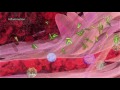 Inflammation
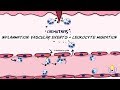 Inflammation: Vascular events and leukocyte migration
 Tissue response to inflammation
 How does your immune system work? - Emma Bryce
 Inflammation - Vascular Events
 B Cells vs T Cells | B Lymphocytes vs T Lymphocytes - Adaptive Immunity - Mechanism
 Inflammation - causes, symptoms, diagnosis, treatment, pathology
 Inflammation - Inflammatory Response - What Is Inflammation In The Body?