Renal Cell Carcinoma for USMLE
Renal Cell Carcinoma Anatomy, Epidemiology, Etiology, Clinical Signs and Symptoms, Treatment and Management. Handwritten, full lecture for medical students taking USMLE.
Renal Cell Carcinomas make up 90-95% of kidney neoplasms.
ETIOLOGY of Renal Cell Carcinoma
Smoking is the largest risk factor. Obesity and Hyppertension is a known risk factor for Renal Cell Carcinoma in Women. Occupational Exposure such as Trichloroethylene, Benzine, Herbicides, Vinyl Chloride. Drugs associated with Renal Cell Carcinoma (phenacitin). Long term dialysis increases the risk of cystic Diseases which increase risk of renal cell carcinoma.
Von Hipel Lindau - Loss of 3p increases HIF which increases angiogenesis. Also increase risk of pheochromocytoma, pancreatic cysts/islet cell tumors, retinal angiomas, CNS hemangioblastomas.
Hereditary Papillary Renal Carcinoma - MET Gene mutation of tyrosine kinase domain and will have bilateral multifocal papillary renal cell carcinoma.
Burt-Hogg-Dube Syndrome - Bilateral Multifocal oncocytoma which has a better prognosis. Also may cause pulmonary and colonic tumors.
CLINICAL SIGNS AND SYMPTOMS of Renal Cell Carcinoma.
The three most common presenting signs and symptoms is flank pain, hematuria, flank mass. A large percentage of patients may be asymptomatic. Patients with renal cell carcinoma may also have wieght loss, varicocele, malaise, fever.
Paraneoplastic syndromes are very common in renal cell carcinoma. Increase EPO may lead to polycythemia, Renin production may lead to hypetension. Finally may also have hypercalcemia, polyneuropathy.
Shauffer Syndrome - Non-metastatic Hepatic Dysfunction and therefore it is important to monitor liver function, even if no metastasis has occurred.
Metastasis to Lungs (45%), Soft tissue and Liver.
Work Up for Renal Cell carcinoma
Labs - Urinalysis, CBC, Electrolytes, Renal Profile, LFT (AST/ALT) and Serum Calcium.
Imaging - CT scan is the imaging of choice and can identify the tumor and rule out cystic mass. Also allows visualtion of Lymph nodes, Renal Vein, IVC and helps rule out angiolipoma.
For staging abdominal ;pelvic CT with or without contrast. Chest X-ray and Brain MRI.
Histology
Clear Cell Carcinoma - 75%, lipid/glycogen
Chromphilic - Bilateral mulftifocal
Chromophobic - Large polygonal Cells
Oncocytoma - Rarely metastasize
Collecting Tubules
STAGING OF Renal Cell Carcinoma
Stage 1 - Within the kidney and less than 7cm.
Stage 2 - Within the kidney and greater than 7cm.
Stage 3 - Invasion Renal Vein and Inferior Vena Cava or Adrenal Gland, but does not invade Gerota's Fascia
Stage 4 - Extends below Gerota's Fascia, invade nearby lymph nodes and metastasis to organs.
MANAGEMENT of Renal Cell Carcinoma
Surgical
Partial nephrectomy for stage 1 and sometimes stage 2
Radical Nephrectomy - remove complete removal of Gerota's fascia, Removal of kidney with adrenal gland, dissect enlarge lymph nodes.
Palliative Nephrectomy - remove kidney to alleviate pain, polycythemia and hypertension.
Adjuvant Treatment for Renal Cell Carcinoma
Biologic Response Mediators - IL2 (activates T Cell and NK), IFN
Molecular Targeting - Suritinib, Bevacizumab, Dazopomib, Temsirolimu, Sorafenib.
Chemotherapy - 5 floururacil, Vinblastine, Paclitaxel, Caboplatin, Ifosfamide, Gemcitabine.
Radiation - Renal Cell Carcinoma is not sensitive to radiation but the brain metastasize are sensitive.
Renal Artery Embolization inject ethanol or gelatin sponge pledgets in artery feeding tumor to help kill off the tumor. Also done palliative for non-surgical patient.
Видео Renal Cell Carcinoma for USMLE канала the study spot
Показать
Комментарии отсутствуют
Информация о видео
Другие видео канала
 Bladder Cancer for USMLE Step 2
 Renal Masses: Inpatient and Outpatient Evaluations (and Updates in Renal Cell Carcinoma)
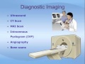 Understanding Kidney Cancer
 Testicular cancer- causes, symptoms, diagnosis, treatment, pathology
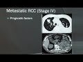 Renal Cell Carcinoma
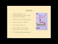 Chronic Kidney Disease - CRASH! Medical Review Series
 Wilm's Tumor
 Bladder Cancer - Overview (types, pathophysiology, diagnosis, treatment)
 RENAL CELL CARCINOMA - How To DIAGNOSE & TREAT /HEMATURIA
 Current Treatment of advanced renal cell carcinoma, Christian Kollmannsberger, MD
 The Kidney and Kidney Cancers | UCLA Urology
 Autosomal Dominant Polycystic Kidney Disease (ADPKD) - causes, pathophysiology, diagnosis, treatment
 Benign Prostate Hyperplasia (BPH) and Prostate Cancer for USMLE Step 2
 Urinary/Kidney Stones - Overview (signs and symptoms, risk factors, pathophysiology, treatment)
 12DaysinMarch, Testicular Tumors for USMLE Step One
 Avoiding Unnecessary Treatment of the Small Renal Mass | Brian Shuch, MD | UCLAMDChat
 Recent Advances in Renal Cell Carcinoma
 Dr.Pritesh Discusses outline of 'KIDNEY AND URETER'
 Essentials in Oncologic Imaging: What Radiologists Need to Know About Renal Cancer Pt. 2
 Renal tuberculosis