
- Популярные видео
- Авто
- Видео-блоги
- ДТП, аварии
- Для маленьких
- Еда, напитки
- Животные
- Закон и право
- Знаменитости
- Игры
- Искусство
- Комедии
- Красота, мода
- Кулинария, рецепты
- Люди
- Мото
- Музыка
- Мультфильмы
- Наука, технологии
- Новости
- Образование
- Политика
- Праздники
- Приколы
- Природа
- Происшествия
- Путешествия
- Развлечения
- Ржач
- Семья
- Сериалы
- Спорт
- Стиль жизни
- ТВ передачи
- Танцы
- Технологии
- Товары
- Ужасы
- Фильмы
- Шоу-бизнес
- Юмор
Traumatic pneumothorax and pneumomediastinum with subcutaneous emphysema #shorts
Traumatic pneumothorax and pneumomediastinum with subcutaneous emphysema
X-ray chest (PA view)
Extensive subcutaneous emphysema is seen as linear lucencies throughout the soft tissues of the thorax, neck, and upper abdomen (examples indicated by green overlay). A small residual right pneumothorax (arrowheads) is visible and pneumomediastinum is seen along the trachea, main bronchi, and cardiac silhouette (red overlay and arrows). A chest tube (yellow overlay) is in place, with its tip in the apex of the right hemithorax.
Pneumomediastinum and pneumothorax can result from blunt or penetrating thoracic injury and various other conditions (e.g., lung disease, intestinal perforation, medical procedure). Air that escapes from the lungs, airways, or bowel can track into the mediastinal and subcutaneous soft tissues.
Видео Traumatic pneumothorax and pneumomediastinum with subcutaneous emphysema #shorts канала Ultrasound@
X-ray chest (PA view)
Extensive subcutaneous emphysema is seen as linear lucencies throughout the soft tissues of the thorax, neck, and upper abdomen (examples indicated by green overlay). A small residual right pneumothorax (arrowheads) is visible and pneumomediastinum is seen along the trachea, main bronchi, and cardiac silhouette (red overlay and arrows). A chest tube (yellow overlay) is in place, with its tip in the apex of the right hemithorax.
Pneumomediastinum and pneumothorax can result from blunt or penetrating thoracic injury and various other conditions (e.g., lung disease, intestinal perforation, medical procedure). Air that escapes from the lungs, airways, or bowel can track into the mediastinal and subcutaneous soft tissues.
Видео Traumatic pneumothorax and pneumomediastinum with subcutaneous emphysema #shorts канала Ultrasound@
Комментарии отсутствуют
Информация о видео
3 июня 2025 г. 18:02:47
00:01:03
Другие видео канала
 #denguefever
 Between boy and girl #cute #love #memes #funny #dop2
 #gallbladder
 Finding baby boy and girl of obstetric ultrasound #girl #boy #ultrasound #obstetricsandgynecology
 How to measurement of Benign prostatic hyperplasia, #ultrasound #abdominal
 He or She ? Obstetrics ultrasound findings #shorts #pregnant #quiz #obstetrician #ultrasoundresult
 #Placenta
 #endometriosis
 Aspect échographie Compatible avec un fibrome intra -mural utérin .#pregnancy #ultrasound
 A gender between boy and girl ultrasound , #pregnancy #ultrasound #boy #girl
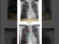 Tension pneumothorax #pneumothorax
 #Hydroureteronephrosis
 learning, what are difference between baby boy and girl ultrasound #ultrasound
 Boy or girl #shorts #pregnant #baby #babygirl #babyboy #funny
 #Fetus Anatomy
 Acute appendicitis #shorts
 A baby Thoracopagus showing , abnormally baby twins #pregnancy #ultrasound #twins #thoracopgaus
 Garden classification of femoral neck fractures
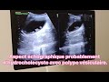 Aspect échographique probablement d'hydrocholecyste avec polype vésiculaire.
 13w6d #pregnant #boyvsgirl #nubia #facts #quiz #vocabulary #english #englishlanguage
 Gender reveal #boyvsgirl #shorts
