Ankle Ligaments Anatomy - Everything You Need To Know - Dr. Nabil Ebraheim
Dr. Ebraheim’s educational animated video describes the anatomy of the ankle ligaments.
The ankle joint is made of three bones, the tibia, the fibula and the talus. The tibia is the major bone of the lower leg which bears the majority of the body weight. At the angle, the bump of the tibia forms the medial malleolus. The fibula is the smaller of the two bone of the leg. The lateral end of the fibula forms the lateral malleolus. In the ankle joint the talus articulates with the tibia. The talus is involved in multiple movements of the foot.
There are ligaments in the ankle that provide connections between the bones. Injury to any of these ligaments may occur when the foot twists, rolls or turns beyond its normal motion. An ankle sprain is a common injury that occurs in sports as basketball and soccer.
The deltoid ligament is on the medial side. It is formed of four parts: anterior tibiotalar part, tibionavicular part, tibiocalcaneal part and the posterior tibiotalar. The superficial deltoid arises from the anterior colliculus. The deep deltoid arises form the posterior colliculus and the intercollicular groove. The deltoid ligament is the main stabilizer of the ankle joint during the stance phase. The deltoid ligament is rarely injured by itself and it is usually associated with fractures.
There are 3 lateral ligaments of the ankle joint:
The anterior talofibular ligament (weakest): origin: 10 mm proximal to the tip of the fibula. Extends from the anterior inferior border of the fibula to the neck of the talus.
The posterior talofibular ligament ( strongest): origin from the posterior border of the fibula. Inserts into posterolateral tubercle of the talus
Calcaneofibular ligament: origin anterior border of the fibula 1 cm proximal to the distal tip. Inserts into the calcaneus distal to the subtalar joint and deep to the peroneal tendon sheath. The lateral ligaments are the most commonly injured ligaments in the ankle.
The ligament of the syndesmosis
•Anterior inferior tibiofibular ligament
•Interosseous ligament
•Posterior inferior tibiofibular ligament
The connection of the tibia and fibula is called the syndesmosis.
High ankle sprain = syndesmosis injury 5-10%. Injury of the ligaments above the ankle.
Become a friend on facebook:
http://www.facebook.com/drebraheim
Follow me on twitter:
https://twitter.com/#!/DrEbraheim_UTMC
Видео Ankle Ligaments Anatomy - Everything You Need To Know - Dr. Nabil Ebraheim канала nabil ebraheim
Показать
Комментарии отсутствуют
Информация о видео
Другие видео канала
 Tennis Elbow Muscle, Extensor Carpi Radialis Brevis -Everything You Need To Know -Dr. Nabil Ebraheim
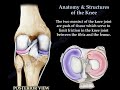 Anatomy & Structures Of The Knee , knee pain
 Anatomy Of The Peroneal Muscles Lower Leg - Everything You Need To Know - Dr. Nabil Ebraheim
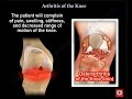 Knee Pain , knee Arthritis. what is knee arthritis ?
 Anatomy Of The Psoas muscle #shorts
 Johnny Widmer Marine Artist & Fisherman - Support His Art Contest
 Compartment Syndrome Of The Thigh - Everything You Need To Know - Dr. Nabil Ebraheim
 Illustrated anatomy of the piriformis, its relationship to the sciatic nerve, piriformis syndrome
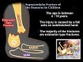 Supracondylar Fracture Of The Humerus In Children - Everything You Need To Know - Dr. Nabil Ebraheim
 SLAP Tear - Everything You Need To Know - Dr. Nabil Ebraheim
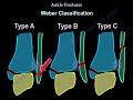 Ankle Fractures types symptoms and treatment
 Gluteus Maximus Gait - Everything You Need To Know - Dr. Nabil Ebraheim
 Reiter's Syndrome Reactive Arthritis - Everything You Need To Know - Dr. Nabil Ebraheim
 Trabecular Pattern of the Proximal Femur - Everything You Need To Know - Dr. Nabil Ebraheim
 Exam of Upper Extremity nerves post operatively - Everything You Need To Know - Dr. Nabil Ebraheim
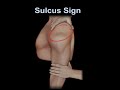 Sulcus Sign, shoulder instability
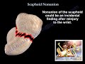 Scaphoid Nonunion - Everything You Need To Know - Dr. Nabil Ebraheim
 Camptodactyly - Everything You Need To Know - Dr. Nabil Ebraheim
 Compartment Syndrome Thigh Fasciotomy - Everything You Need To Know - Dr. Nabil Ebraheim
 Anatomy Of The Subscapularis Muscle - Everything You Need To Know - Dr. Nabil Ebraheim
 Peroneus Tertius Muscle - Everything You Need To Know - Dr. Nabil Ebraheim