Identifying lesion/stenosis severity during coronary angiography (and PCI)
In this video, you'll learn to identify non-obstructive coronary artery disease (CAD) vs severe /obstructive CAD. If the lesion is intermediate, you will learn ways in which we can further assess significance later in PCI Essentials course (see below). We'll talk about 'eye-ball' assessment or QCA and introduce other modalities of imaging.
Perform your own percutaneous coronary interventions (PCI) with our Percutaneous Coronary Intervention Essentials course. You’ll cover essential PCI equipment, lesion assessment, troubleshooting, and patient monitoring so you can perform your own procedures under the supervision of an experienced cardiologist. This course will debunk common myths surrounding PCI patient selection so you can offer it to more patients who need it. Get your FREE trial account and start chapter 1 NOW!
Course: https://www.medmastery.com/course/percutaneous-coronary-intervention-essentials
Free Account: www.medmastery.com/user/register
Видео Identifying lesion/stenosis severity during coronary angiography (and PCI) канала Medmastery
Показать
Комментарии отсутствуют
Информация о видео
Другие видео канала
 IVUS and FFR (Colin M. Barker, MD) Saturday, August 20, 2016
 Using a pressure wire in percutaneous coronary interventions (PCI)
 Scoring coronary artery calcium (CAC) with cardiac CT
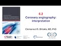 6.2 Manual of PCI - How to interpret the coronary angiogram
 Coronary anatomy refresher
 The left coronary artery - A walk-through
 Case 21: Manual of non-CTO interventions: acute stent thrombosis
 Video case study: IVUS and iFR-guided PCI of 71-year old male with diffuse coronary artery disease
 OCT-guided PCI: step-by-step_Shlofmitz_Coronary On Demand
 IVUS and FFR (Syed Gilani, MD)
 Coronary angiography - image intensifier basics and nomenclature
 Case 68 - Manual of PCI - branch ostial lesion
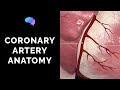 Coronary Artery Anatomy (3D Anatomy Tutorial)
 22.2 In-stent restenosis: Manual of PCI
 Diffuse disease and non dilatable lesion.
 Basics of angiographic views during left heart catheterization
 Coronary angiography. Lesson 1: coronary artery anatomy
 Case 44: Manual of non-CTO coronary interventions - wiring in STEMI
 Coronary Artery Disease - Left coronary artery stenosis
 15.2 Branch ostial lesions - Manual of PCI