PreOp® Patient Education Anti-Reflux Laparoscopy Surgery 2
http://bit.ly/PreOpFacebook or http://bit.ly/PreOpTwitter - Patient Education - http://www.PreOp.com
Patient Education Company
After allowing a few minutes for the anesthetic to take effect...
a small incision is made above the umbilicus;
then, a hollow needle will be inserted through the abdominal wall.
And the abdomen will be inflated with carbon dioxide.
An umbilical port is created for the laparoscope.
Four more incisions will be made, with care taken to keep the openings as small as possible.
Once in place, the laparoscope will provide video images,
so the surgeon can insert the instruments used to locate and pull back the liver...
in order to see the upper part of the stomach.
Then, the surgeon cuts away the tissue that connects the liver and the stomach..
Then the surgeon divides and separates the arteries that supply blood to the top of the stomach.
After freeing the stomach from the spleen,
your doctor wraps the upper portion of the stomach around the esophagus and sutures it into place.
A rubber tube is placed in the esophagus to keep the wrap from becoming too tight.
All of the instruments are withdrawn...
the carbon dioxide is allowed to escape...
the muscle layers and other tissues are sewn together and the skin is closed with sutures or staples.
Finally, sterile dressings are applied.
Patient Education Company
Видео PreOp® Patient Education Anti-Reflux Laparoscopy Surgery 2 канала PreOp.com Patient Engagement - Patient Education
Patient Education Company
After allowing a few minutes for the anesthetic to take effect...
a small incision is made above the umbilicus;
then, a hollow needle will be inserted through the abdominal wall.
And the abdomen will be inflated with carbon dioxide.
An umbilical port is created for the laparoscope.
Four more incisions will be made, with care taken to keep the openings as small as possible.
Once in place, the laparoscope will provide video images,
so the surgeon can insert the instruments used to locate and pull back the liver...
in order to see the upper part of the stomach.
Then, the surgeon cuts away the tissue that connects the liver and the stomach..
Then the surgeon divides and separates the arteries that supply blood to the top of the stomach.
After freeing the stomach from the spleen,
your doctor wraps the upper portion of the stomach around the esophagus and sutures it into place.
A rubber tube is placed in the esophagus to keep the wrap from becoming too tight.
All of the instruments are withdrawn...
the carbon dioxide is allowed to escape...
the muscle layers and other tissues are sewn together and the skin is closed with sutures or staples.
Finally, sterile dressings are applied.
Patient Education Company
Видео PreOp® Patient Education Anti-Reflux Laparoscopy Surgery 2 канала PreOp.com Patient Engagement - Patient Education
Показать
Комментарии отсутствуют
Информация о видео
15 декабря 2009 г. 10:31:18
00:01:40
Другие видео канала
 A Virtual Reality tour inside the Heart
 360° VR Bone Resorption | Nucleus Medical Media
 VR Patient Experience COMPRESSED
 Ciclo menstrual
 insta evo seagull 3d 180 poop on beach
 #Bl #boylove #Mpreg #wattpad Unboxing buku novel Jamaludin. (cowok hamil)
 LOOK AT MY BELLY (180 degree VR)
 3 PM | Ghantaravam | News Headlines | 24th June 2021 | ETV Telangana
 Multivrs 360° Medical Experience
 BOLD Faces #2
 5 Hábitos ruins de quem programa
 G.z ToDay
 360 Degree Tour of MedAire's MedLink In-Flight Emergency Response Center
 TURP Transurethral Resection Prostate - PreOp® Patient Education & Patient Engagement #shorts
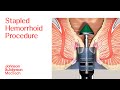 PPH Procedure Animation | Stapled Hemorrhoid Surgery
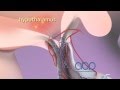 Female Reproductive System
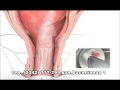 Лікування геморою за 1 візит в "Оксфорд Медікал Прикарпаття"
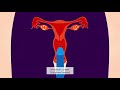 Contraception in Tamil
 Altis surgery animation
 BS. LÊ THỊ NHÀI CHIA SẺ : QUY TRÌNH THẨM MỸ CẮT MÔI BÉ