Ultrasound Video showing Endometrial hyperplasia in 33 years old female.
This video shows Endometrial hyperplasia in 33 years old female.
Endometrial hyperplasia is a histological diagnosis often made after sampling an endometrium that appears thickened on pelvic ultrasound. It is defined as an irregular proliferation of endometrial glands with an increased ratio of the gland to the stroma.
The endometrial hyperplasia has a cystic lace-like appearance on ultrasound. Endometrial polyps manifest as focal areas of endometrial thickening, and the stalk of the polyp may be seen if sufficient fluid is present in the endometrial cavity.
Endometrial hyperplasia is a condition in which the endometrium (the lining of the uterus) becomes abnormally thick. Although endometrial hyperplasia is not cancer, it can lead to uterine cancer in some women. Endometrial hyperplasia is usually caused by an excess of estrogen without progesterone (female hormones).
Ultrasound is often one of the first tests used to look at the uterus, ovaries, and fallopian tubes in women with possible gynecologic problems. Images from the TVUS can be used to see if the uterus contains a mass (tumor), or if the endometrium is thicker than usual, which can be a sign of endometrial cancer.
Endometrial hyperplasia is a noncancerous (benign) condition where the lining of the womb becomes thicker. There is a higher risk of developing womb cancer if a female has this thickening, especially if the extra lining cells are abnormal.
Among postmenopausal women with vaginal bleeding, the endometrial thickness of less than 5 mm is generally considered normal, while thicknesses 5 mm or more is considered abnormal.
The most common signs of excessive endometrial thickness include:
Bleeding after menopause.
Extremely heavy or long-lasting bleeding during menstruation.
Irregular menstrual cycles that last less than 3 weeks or longer than 38 days.
spotting between periods.
Endometrial hyperplasia is a histological diagnosis often made after sampling an endometrium that appears thickened on pelvic ultrasound. It is defined as an irregular proliferation of endometrial glands with an increased ratio of the gland to the stroma.
Atypical hyperplasia can turn into cancer of the womb. 20 years after diagnosis, around 28 out of every 100 women diagnosed with atypical hyperplasia will develop cancer of the womb.
Your doctor can perform an exam and tests to diagnose the main condition. A transvaginal ultrasound measures the endometrium. A thick layer can indicate endometrial hyperplasia. Biopsy of the endometrium cells is taken to determine if cancer is present.
In one study, among women 18–90 years the overall incidence of endometrial hyperplasia was 133 per 100,000 woman-years, was most common in women ages 50–54, and was rarely observed in women under 30. Simple and complex hyperplasia incidences peaked in women ages 50–54.
Early warning signs of uterine cancer
Unusual vaginal discharge that does not have signs of blood.
Difficult or painful urination.
Pain during intercourse.
Pain and/or a mass in the pelvic area.
Unintentional weight loss.
The most common treatment is progestin. This can be taken in several forms, including the pill, shot, vaginal cream, or an intrauterine device. Atypical types of endometrial hyperplasia, especially complex, increase your risk of getting cancer. If you have these types, you might consider a hysterectomy.
Видео Ultrasound Video showing Endometrial hyperplasia in 33 years old female. канала Saeed Ahmad
Endometrial hyperplasia is a histological diagnosis often made after sampling an endometrium that appears thickened on pelvic ultrasound. It is defined as an irregular proliferation of endometrial glands with an increased ratio of the gland to the stroma.
The endometrial hyperplasia has a cystic lace-like appearance on ultrasound. Endometrial polyps manifest as focal areas of endometrial thickening, and the stalk of the polyp may be seen if sufficient fluid is present in the endometrial cavity.
Endometrial hyperplasia is a condition in which the endometrium (the lining of the uterus) becomes abnormally thick. Although endometrial hyperplasia is not cancer, it can lead to uterine cancer in some women. Endometrial hyperplasia is usually caused by an excess of estrogen without progesterone (female hormones).
Ultrasound is often one of the first tests used to look at the uterus, ovaries, and fallopian tubes in women with possible gynecologic problems. Images from the TVUS can be used to see if the uterus contains a mass (tumor), or if the endometrium is thicker than usual, which can be a sign of endometrial cancer.
Endometrial hyperplasia is a noncancerous (benign) condition where the lining of the womb becomes thicker. There is a higher risk of developing womb cancer if a female has this thickening, especially if the extra lining cells are abnormal.
Among postmenopausal women with vaginal bleeding, the endometrial thickness of less than 5 mm is generally considered normal, while thicknesses 5 mm or more is considered abnormal.
The most common signs of excessive endometrial thickness include:
Bleeding after menopause.
Extremely heavy or long-lasting bleeding during menstruation.
Irregular menstrual cycles that last less than 3 weeks or longer than 38 days.
spotting between periods.
Endometrial hyperplasia is a histological diagnosis often made after sampling an endometrium that appears thickened on pelvic ultrasound. It is defined as an irregular proliferation of endometrial glands with an increased ratio of the gland to the stroma.
Atypical hyperplasia can turn into cancer of the womb. 20 years after diagnosis, around 28 out of every 100 women diagnosed with atypical hyperplasia will develop cancer of the womb.
Your doctor can perform an exam and tests to diagnose the main condition. A transvaginal ultrasound measures the endometrium. A thick layer can indicate endometrial hyperplasia. Biopsy of the endometrium cells is taken to determine if cancer is present.
In one study, among women 18–90 years the overall incidence of endometrial hyperplasia was 133 per 100,000 woman-years, was most common in women ages 50–54, and was rarely observed in women under 30. Simple and complex hyperplasia incidences peaked in women ages 50–54.
Early warning signs of uterine cancer
Unusual vaginal discharge that does not have signs of blood.
Difficult or painful urination.
Pain during intercourse.
Pain and/or a mass in the pelvic area.
Unintentional weight loss.
The most common treatment is progestin. This can be taken in several forms, including the pill, shot, vaginal cream, or an intrauterine device. Atypical types of endometrial hyperplasia, especially complex, increase your risk of getting cancer. If you have these types, you might consider a hysterectomy.
Видео Ultrasound Video showing Endometrial hyperplasia in 33 years old female. канала Saeed Ahmad
Показать
Комментарии отсутствуют
Информация о видео
Другие видео канала
 Ultrasound Video showing a case of Endometrial hyperplasia.
 Ultrasound Video showing Three types of Cysts in the same patient.
 Understanding the Endometrium on Ultrasound
 Uterus Ultrasound Normal Vs Abnormal Image Appearances Comparison | Uterine Pathologies USG
 Endometrial cancer - causes, symptoms, diagnosis, treatment, pathology
 What is a 'thickened' endometrium? Should you be worried?
 Best Treatment for #Endometriosis | Womens Health | Dr Rooma Sinha | Apollo Hospitals Hyd
 Uterine Polyp and Ovarian Hemorrhagic Cyst.
 31) Endometrial Hyperplasia: Treatment with the LNG IUD (@dr_dervaitis)
 Endometriosis
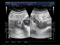 Endometrial hyperplasia after menopause, women aged 54 years.
 Ultrasound Video showing the technique to localize the Inflamed Appendix.
 RCOG GUIDELINE MANAGEMENT OF ENDOMETRIAL HYPERPLASIA
 ENDOMETRIAL HYPERPLASIA
 Endometrial polyp ultrasound
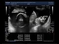 Ultrasound Video showing twin pregnancy with one anencephalic fetus and polyhydromnios.
 Ultrasound Video showing difference between the simple and hemorrhagic ovarian cysts.
 Endometrial hyperplasia - an Osmosis Preview
 Endometrial Cancer - The Hidden Signs
 Hepatic Hydatid Cyst.