- Популярные видео
- Авто
- Видео-блоги
- ДТП, аварии
- Для маленьких
- Еда, напитки
- Животные
- Закон и право
- Знаменитости
- Игры
- Искусство
- Комедии
- Красота, мода
- Кулинария, рецепты
- Люди
- Мото
- Музыка
- Мультфильмы
- Наука, технологии
- Новости
- Образование
- Политика
- Праздники
- Приколы
- Природа
- Происшествия
- Путешествия
- Развлечения
- Ржач
- Семья
- Сериалы
- Спорт
- Стиль жизни
- ТВ передачи
- Танцы
- Технологии
- Товары
- Ужасы
- Фильмы
- Шоу-бизнес
- Юмор
JID February 2024 Cover: SARS-CoV-2 from Skin Lesion to Ultrastructure
COVID-19; SARS-CoV-2; Spike protein; Vasculitis; Immunofluorescence; Electron microscopy; Dermatology
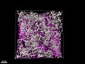
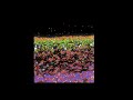
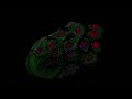
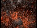
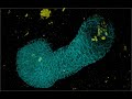
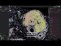
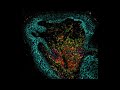
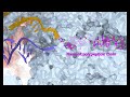
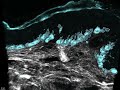
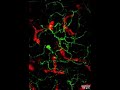
Overview of SARS-CoV-2-Related Vasculitic Skin Lesions: A comprehensive journey from clinical images to ultrastructure. Dermal vascular cell destruction is evident in H&E staining, while electron microscopy reveals the presence of autophagosomes. Immunofluorescence images showcase spike protein depositions in COVID-19-associated autophagosomes of dermal microvascular cells. Staining details for immunofluorescent images: SARS-CoV-2 spikes (blue), LC3B (red), LC3C (green), DAPI (gray).
Video courtesy of Simon Riel, Core Facility for Electron Microscopy, Department of Dermatology, University Hospital Tübingen, Germany.
Видео JID February 2024 Cover: SARS-CoV-2 from Skin Lesion to Ultrastructure канала Journal of Investigative Dermatology (JID)
Комментарии отсутствуют
Информация о видео
12 января 2024 г. 19:50:26
00:00:57
Другие видео канала