Beautiful reconstruction of a mouse dopamine neuron.
The video shows sparse-labelling of a single dopaminergic neuron with a fluorescent protein, in a mouse brain hemisphere, that has been cleared of fat to become translucent. A z-stack of lightsheet imaging reconstructs the entire cell from its soma in the midbrain, via a single axon, to its dopaminergic terminal field in the striatum (~450,000 terminals). Work made possible by a Fixel Chair in Parkinson’s disease to Dr. Matt Farrer
Видео Beautiful reconstruction of a mouse dopamine neuron. канала Benjamin Stecher
Видео Beautiful reconstruction of a mouse dopamine neuron. канала Benjamin Stecher
Показать
Комментарии отсутствуют
Информация о видео
Другие видео канала
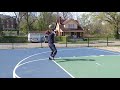 Basketball as Therapy for Parkinson's Disease
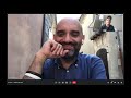 Interview with Prof. Alfonso Fasano on MAID (Medical Assistance In Dying)
 Porridge for Parkinson's talk
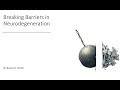 Breaking Barriers in Neurodegeneration Benjamin Stecher June 21, 2019
 Overreacting
 Introduction to Deep Brain Stimulation by: Benjamin Stecher and Gina Lupino
 Olivia Samotus explaining Spinal Cord Stimulation
 DBS sweet spot.
 PD Warrior Presentation With Ben & Alfonso
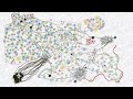 Role of Glia Cells in Health, Aging and Disease
 Interview w/ Prof. Lee Smolin on Parkinson's and getting a Deep Brain Stimulator implanted yesterday
 Behind the Scenes of Cryogenic Electron Microscopy with Prof. Henning Stahlberg
 Wishlist for Neurodegenerative Disease Research
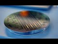 The Future of Brain-Machine Interfaces
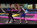 Bouncing around with Ju Domingos
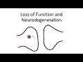 Loss of Function and Neurodegeneration
 How we discovered how to restore memories
 A Few Highlights From the First Programming Session
 The Three Pillars of Deep Brain Stimulation Therapy...and much much more
 Brief Interview with David Ashford Jones on getting DBS at the start of Lockdowns in London.