How to do a Basic Transthoracic Echocardiogram: Transducer Position and Anatomy
"How to Perform a Transthoracic Echocardiographic Study Volume 1: Transducer Position and Anatomy" is an instructional video, offered by ASE, and can be used for professional lectures and offers an interactive section for flexible presentations. The video includes an overview of relevant cardiac anatomy, a step by step presentation of all Transducer Positions, and the sequential transducer movements to acquire standard echo images needed to complete a Transthoracic Echocardiographic Study.
The cardiac anatomy section visualizes relationships of the heart, ribs, axis of the heart, planes of the heart and aorta, chambers, valves and annuli, and blood flow through the heart.
The transducer position menu allows the user to view the anatomical landmarks and the transducer positions for left parasternal, apical, subcostal, suprasternal and right parasternal image acquisition.
The views and imaging menu allows the user to navigate from a particular transducer position menu such as left parasternal long to specific images acquired within that window such as PLAX high depth, PLAX low depth, and PLAX Zoom of the Aortic and Mitral Valves. Each animated loop shows a finder image in the upper left that depicts the transducer position, an illustration of the sectional anatomy of the heart on the left, and the echo image with labels on the right.
Видео How to do a Basic Transthoracic Echocardiogram: Transducer Position and Anatomy канала ASE360
Показать
Комментарии отсутствуют
Информация о видео
Другие видео канала
 Basic Transthoracic Echocardiography (Cardiac Ultrasound) - TTE Made Simple
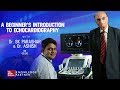 A Beginner's Introduction To Echocardiography | Dr. S.K. Parashar | Echo Masterclass
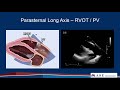 Part 2: Comprehensive TTE in Adults Webinar
 Intro to Echo Parasternal Views.mov
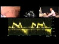 Diastolic Function — A Simple Approach
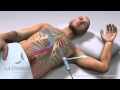 How to obtain: Apical 4 (Four) Chamber Ultrasound View- Training and Techniques - ICU
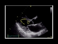 Estimating Ejection Fraction with Point of Care Echo
 Echocardiography Essentials: Mastering the apical four-chamber view (4CV)
 Transthoracic Echo full protocol. Part II: Parasternal View (PLAX , PSAX, RVIT, RVOT, M-Mode)
 Echocardiography Essentials: Mastering the suprasternal view of the aorta
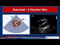 Guidelines for Performing a Comprehensive TTE in Adults Webinar
 Echo BachelorClass - Your introduction to basic echocardiography
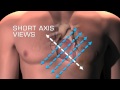 Parasternal Short Axis Views
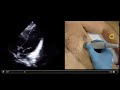 Optimizing Cardiac Views with Ultrasound
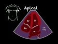 Bedside Ultrasound Basic Cardiac US
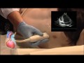 The 2 Chamber View in Echocardiography
 Echocardiography for beginners
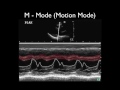 Ultrasound Physics Scanning Modes M Mode
 Basic TTE Video Tutorial
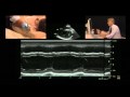 M-Mode Measurement of the Left Ventricle