Anatomy of Middle cranial Fossa
Join this channel to get access to perks:
https://www.youtube.com/channel/UCG5TBPANNSiKf1Dp-R5Dibg/join
Follow on Instagram :- https://www.instagram.com/drgbhanuprakash
Middle cranial fossa
---------------------------------
The middle cranial fossa (latin: fossa cranii media) is a region of the internal cranial base between the anterior and posterior cranial fossae, it lies deeper and is wider than the anterior cranial fossa.
The floor of the middle cranial fossa is formed by the body and greater wings of the sphenoid, the squamous part of the temporal bone, and the anterior surface of the petrous part of the temporal bone.
The boundaries of the middle cranial fossa are formed anteriorly by the lesser wings and part of the body of the sphenoid, posteriorly by the superior borders of the petrous part of the temporal bone and the dorsum sellae of the sphenoid, laterally by the squamous parts of the temporal bones, the parietal bones, and the greater wings of the sphenoid.
Borders
-------------
These are the borders of the middle cranial fossa:
anteromedial: limbus of the sphenoid;
anterolateral: lesser wings and part of the body of the sphenoid;
posteromedial: dorsum sellae of the sphenoid;
posterolateral: superior margin of the petrous part of the temporal bone;
lateral: squamous parts of the temporal and parietal bones;
inferior: body and greater wings of the sphenoid, squamous and petrous parts of the temporal bone.
Contents
---------------
The middle cranial fossa accommodates the following anatomical structures:
pituitary gland,
temporal lobes of the cerebral cortex.
Openings
----------------
There are many openings in the middle cranial fossa connecting it to other parts of the skull, these are the following:
optic canal,
superior orbital fissure,
foramen rotundum,
foramen ovale,
foramen spinosum,
foramen lacerum,
carotid canal,
hiatus for lesser petrosal nerve,
hiatus for greater petrosal nerve.
The pair of optic canals connect the middle cranial fossa with the orbits and transmit the optic nerves and ophthalmic arteries.
The paired superior orbital fissure also connects this fossa with the orbit and transmits the oculomotor, trochlear, ophthalmic and abducens nerves, the ophthalmic veins, and sympathetic fibers.
The foramen rotundum is a paired connection between the middle cranial fossa and the pterygopalatine fossa transmitting the maxillary division of the trigeminal nerve.
The foramen ovale is a paired opening that connects the middle cranial fossa with the external surface of the cranial base and the infratemporal fossa. The foramen ovale transmits the mandibular division of the trigeminal nerve, the lesser petrosal nerve, the accessory meningeal branch of the maxillary artery, and an emissary vein.
The paired foramen spinosum connects the middle cranial fossa with the infratemporal fossa. It transmits the middle meningeal artery, vein and the meningeal branch of the mandibular division of the trigeminal nerve.
The foramen lacerum is a paired opening between the middle cranial fossa and the external surface of the cranial base. The foramen lacerum is filled with cartilage after birth and it transmits the artery and nerve of the pterygoid canal.
The carotid canal and the hiatuses of the lesser and greater petrosal nerves are paired openings in the temporal bone.
The carotid canal connects the middle cranial fossa to the external cranial base and carries the internal carotid artery.
The hiatus for the lesser petrosal nerve transmits the named nerve to the tympanic cavity.
The hiatus for the greater petrosal nerve transmits the greater petrosal nerve and the petrosal branch of the middle meningeal artery from the facial canal to the middle cranial fossa.
#anatomyofmiddlecranialfossa #middlecranialfossa #middlecranialfossaanatomy #cranialfossaanatomy #cranialfossa #anatomy #usmlestep1 #usmle #nationalexittest #mbbsanatomy
Видео Anatomy of Middle cranial Fossa канала Dr.G Bhanu Prakash Animated Medical Videos
Показать
Комментарии отсутствуют
Информация о видео
7 октября 2020 г. 18:30:15
00:16:15
Другие видео канала
 Anatomy of Posterior cranial Fossa
 Anatomy of Anterior cranial Fossa
 TEMPORAL BONE
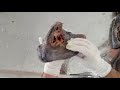 LUNGS PART-1 GENERAL FEATURES AND SIDE DETERMINATION - BY DR MITESH DAVE
 NORMA LATERALIS
 ANATOMY OF STOMACH - BY DR MITESH DAVE
 Dural Folds | Falx cerebri | Tentorium cerebelli | Falx cerebelli | Diaphragma sella | Attachments |
 BONES OF THE SKULL - LEARN IN 4 MINUTES
 Anatomy of the Skull: Norma basalis ( Anterior part , Middle part and Posterior part )
 Middle Cranial Fossa | Skull Anatomy
 SKELETAL SYSTEM ANATOMY: Inferior aspect of the human skull
 Skull bones, sutures and landmarks
 SAGITTAL SECTION OF HEAD & NECK PART-1 : PHARYNX AND RELATED STRUCTURES - BY DR MITESH DAVE
 INTERIOR OF CRANIUM - 2/3
 Posterior Cranial Fossa | Skull Anatomy
 MANDIBLE - GENERAL FEATURES & ATTACHMENTS
 Skull Osteology - Cranial Cavity Anatomy
 HIP BONE | BONES OF LOWER LIMB | ANATOMY | SIMPLIFIED ✔
 Anterior Cranial Fossa | Skull Anatomy
 Anatomy of the Skull : Norma Lateralis