Experiences in Python for Medical Image Analysis; SciPy 2013 Presentation
Authors: Warner, Joshua, Mayo Clinic Department of Biomedical Engineering
Track: Medical Imaging
Upon entering graduate school and selecting radiology informatics as my topic of study, a broad survey of open source options for scientific work was conducted. There were three main criteria:
robust numerical and scientific capability,
strong user community with continuing updates and long term support, and
ease of use for students transitioning from other languages.
Among several strong options that satisfied criteria #1, Python with NumPy and SciPy was the clear winner due to the latter two criteria.
My work focuses on supervised segmentation of soft-tissue abdominal MRI images, extracting novel image features from these segmented regions of interest, and applying machine learning techniques to evaluate features for predictive ability. This presentation will provide an overview of the key computational tasks required for this work, and outline the challenges facing a medical image researcher using Python. Most notably, medical image volumes are rarely isotropic, yet often algorithms for 3-D NumPy arrays inherently assume isotropic sampling. Thus, generalizing or extending various algorithms to handle anisotropic rectangular sampled data is necessary. Our improvements to one such algorithm were recently contributed back to the community, and are presently incorporated in the random walker segmentation algorithm in Scikit-Image.
Another significant challenge is visualization of algorithm output for large volumetric datasets. An extensible tool we call volview was developed, allowing fast visualization of an entire volume and an arbitrary number of colored, alpha-blended overlays, combining the abilities of NumPy, Pyglet, and PygArrayImage. This improved speed and quality of algorithm development, and facilitated review of our results by clinicians.
Видео Experiences in Python for Medical Image Analysis; SciPy 2013 Presentation канала Enthought
Показать
Комментарии отсутствуют
Информация о видео
Другие видео канала
 Deep Learning in Medical Imaging - Ben Glocker, Imperial College London
 AI in Radiology at Stanford: Rise of the Machines
 Xarray for Scalable Scientific Data Analysis | SciPy 2020 | Hamman, Abernathey, Cherian, Hoyer
 Parallel and Distributed Computing in Python with Dask | SciPy 2020 | Bourbeau, McCarty, Pothina
 Medical Image Analysis
 Daniel Chen: Cleaning and Tidying Data in Pandas | PyData DC 2018
 Machine Learning For Medical Image Analysis - How It Works
 Image Analysis in Python with SciPy and scikit-image | SciPy 2018 Tutorial | Stefan van der Walt
 Learn Python through Data Processing in Pandas Tutorial | SciPy 2020 | Daniel Chen
 Python Tutorial: Make images come alive with scikit-image
 16 - Understanding digital images for Python processing
 Reading Files - 3D Convolutional Neural Network w/ Kaggle and 3D medical imaging p.2
 ITK The Insight Segmentation and Registration Toolkit | SciPy 2018 | Matthew McCormick
 MACHINE LEARNING and AUGMENTED REALITY
 Introduction to Conda for (Data) Scientists Tutorial | SciPy 2020 | David Pugh
 Spatial Data Analysis with PySAL Tutorial | SciPy 2020 | Sergio Rey and Elijah Knaap
 Jupyter Interactive Widget Ecosystem Tutorial | SciPy 2020 | Craig, Renou, Dafna, Bektas
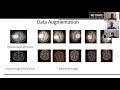 Brain Tumor Detection using Convolutional Neural Network
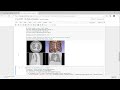 Medical image processing in your web browser using Jupyter notebooks and 3D Slicer
 Template Matching - OpenCV with Python for Image and Video Analysis 11