How do MRI, PET and CAT scans work?
Major advancements in the field of medical imaging now allow healthcare providers to see a clear visual representation of the situation inside a person’s body. This video discusses three of the most commonly used medical imaging techniques: MRI, PET and CAT scans. In three hypothetical scenarios, the video compares and contrasts the three methods in terms of their advantages to the specific medical situation and risks to the patients.
An MRI creates a clear image of the body without posing a serious risk to the patient due to its lack of ionizing radiation.
The clarity and detail of a CAT scan is a focal point when comparing these three techniques, despite its increased risk of cancer due to the ionizing radiation it exposes the patient to. Due to its 3-dimension cross-sectional pictures, CAT scans can be used in a great range of medical complications including the detection of cancer.
By using radioactive tracers, a PET scan differs from the first two techniques in its ability to provide information about organ function rather than just structure. It provides valuable information about functions such as glucose metabolism, oxygen use and blood flow. The PET scan can also provide information at a cellular level.
Finally, the video presents the steps that patients should take to prepare for these tests such as mentioning any previous medical conditions, pregnancy and the presence of metal anywhere on or inside the body which could hinder the results, especially in the MRI and CAT scan.
Video by Zubair Banoor, Jaskeerat Kaur, Sina Nastarani, Kritika Seth and Monica Persaud
Copyright McMaster University 2016
Видео How do MRI, PET and CAT scans work? канала Demystifying Medicine
Показать
Комментарии отсутствуют
Информация о видео
Другие видео канала
 What’s the Difference Between an X-ray, MRI and a CT? | Medical Advice with Doctor ER
 The most important lesson from 83,000 brain scans | Daniel Amen | TEDxOrangeCoast
 How do brain scans work? - John Borghi and Elizabeth Waters
 How does a PET scan work?
 Positron Emission Tomography | PET
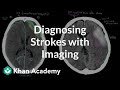 Diagnosing strokes with imaging CT, MRI, and Angiography | NCLEX-RN | Khan Academy
 How does MRI work? Jerome Maller explains
 Xray, CT, MRI differences
 What's The Difference Between An X-Ray, CT Scan and MRI? Which Is Best For Herniated Disc?
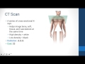 Intro to Clinical Imaging
 How X-rays see through your skin - Ge Wang
 PET-Imaging
 2-Minute Neuroscience: Neuroimaging
 MRI: Basic Physics & a Brief History
 7 Differences between a CT and an MRI scan
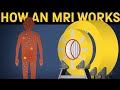 How does an MRI machine work?
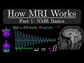 How MRI Works - Part 1 - NMR Basics
 Magnetic Resonance Imaging (MRI)
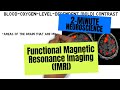 2-Minute Neuroscience: Functional Magnetic Resonance Imaging (fMRI)
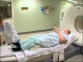 PET (Positron Emission Tomography) scan: What to expect